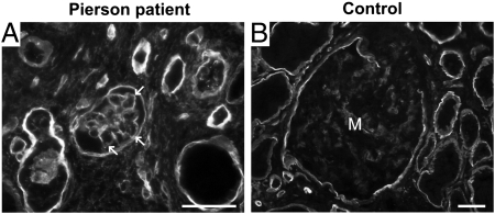
Fig. 2.
Increased Lamβ1 in the GBM in human Pierson syndrome. Immunofluorescence analysis of Lamβ1 in human kidney sections. A 3-mo-old Pierson syndrome patient's specimen (A) shows linear staining for Lamβ1 in the GBM (arrows), whereas a normal adult control (B) shows weak mesangial staining and the absence of staining in the GBM. (Scale bars, 50 μm.)

