Fig. 4.
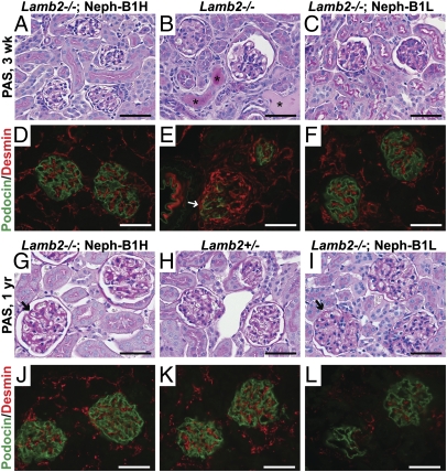
Histological analysis reveals maintenance of podocyte phenotype in Lamb2−/−; Neph-B1 kidneys. (A–C and G–I) PAS staining of kidneys from 3-wk-old (A–C) and 1 y-old (G–I) mice. Asterisks in B indicate protein casts in tubules of a nephrotic Lamb2−/− mouse, which were not observed in Lamb2−/−; Neph-B1 mice (A and C). Arrows in G and I indicate thickening of the GBM at 1 y in Lamb2−/−; Neph-B1 mice. (D–F and J–L) Immunofluorescence analysis of podocin (green) and desmin (red) in kidney sections from 3-wk-old (D–F) and 1-y-old (J–L) mice. Arrow in E indicates an injured podocyte expressing both podocin and desmin; desmin is confined to mesangial cells in normal glomeruli, as observed in the Lamb2−/−; Neph-B1 and Lamb2+/− panels. (Scale bars, 50 μm.)

