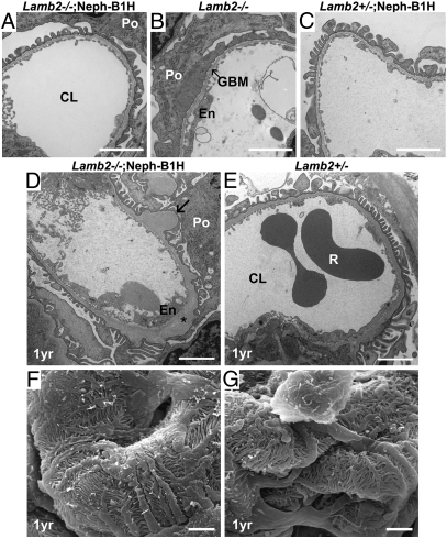
Fig. 6.
Ultrastructural analysis of glomerular capillary walls. (A–E) Transmission electron micrographs of glomerular capillary loops from 3-wk-old (A–C) and from 1-y-old (D and E) mice. Note that the severe podocyte foot process effacement observed in Lamb2−/− mice (B) was not observed in young or old Lamb2−/−; Neph-B1 mice (A and D). Arrow in D indicates segmental thickening of the GBM. Asterisk indicates electron lucent areas in the expanded lamina densa. (F and G) Scanning electron micrographs of glomeruli from 1-y-old mice. Podocyte foot processes in Lamb2−/−; Neph-B1 mice were intact. (Scale bar, 2 μm.) Po, podocyte; CL, capillary lumen; En, endothelial cell; R, red blood cell.

