Fig. 1.
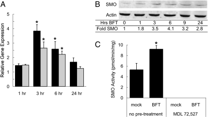
BFT induces SMO in human colon epithelial cell lines. T84 and/or HT29/c1 cells were treated with 5 nM purified, recombinant BFT for the indicated time points. (A) SMO gene expression in T84 (black bars) and HT29/c1 (gray bars) cells was quantified by qRT-PCR as described in Materials and Methods and data from three independent experiments analyzed in triplicate are presented (mean + SEM). Statistically significant (*P < 0.05 by t test) increase in SMO expression vs. untreated cells at the indicated time point. (B) HT29/c1 total cellular lysates were analyzed via quantitative fluorescent Western blotting as described in Materials and Methods and SMO protein levels were normalized to β-actin (representative of two independent experiments). (C) HT29/c1 cells were exposed to BFT for 6 h and SMO and APAO enzyme activity (pmol/min/mg protein) were determined (mean of two independent experiments analyzed in triplicate; *P < 0.05 by t test). Pretreatment overnight with MDL 72527 at concentrations of 10, 25, 50, or 100 μM rendered SMO activity undetectable. APAO enzyme activity was not detected in any samples.

