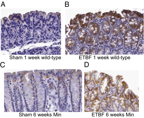
Fig. 3.
ETBF-infected mice exhibit regions of intense SMO staining. C57BL/6 mice were sham or ETBF inoculated and killed after 1 wk (wild type) or 6 wk (Min). Rolled colon tissues were fixed, embedded, and analyzed by immunohistochemistry using specific antisera against SMO. Representative images are displayed from (A) wild type, sham, 1 wk; (B) wild type, ETBF, 1 wk; (C) Min, sham, 6 wk; (D) Min, ETBF, 6 wk. Sham mice had regions of mild-to-moderate apical SMO staining. ETBF-infected mice contained regions of intense SMO staining that extended deeper into the crypts.

