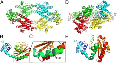
Fig. 1.
The crystal structure of TtgV. (A) TtgV tetramer arrangement. Each monomer is colored differently. (B) Ribbon representation of TtgV monomer colored from N terminus (blue) to C terminus (red). (C) Proposed interaction between the lateral side chains of arginine 98 and aspartic acid 102 (Fig. S1 for further details). (D) TtgV tetramer bound to its target operator. (E) Detail of the kink in the connecting α-helix when TtgV is bound to its target operator. Other details can be found in Lu et al. (10).

