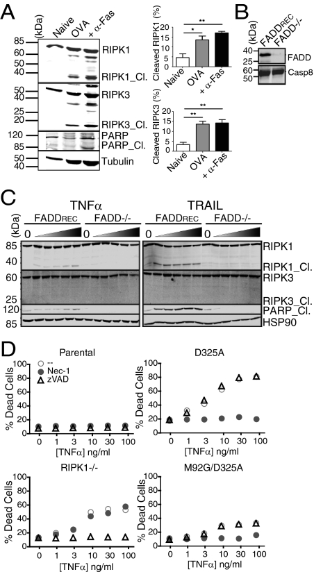
Fig. 5.
RIPK1/RIPK3 cleavage following TCR vs. DR ligation. (A) OT-I T cells (27) were stimulated with OVA peptide for 72 h and stimulated without or with α-Fas or left untreated (naive); immunoblots were hybridized with α-RIPK1 and α-RIPK3 to detect processing. Graphs represent percent RIPK1/3 cleavage (*P < 0.05 and **P < 0.01). (B) Reconstitution of FADD-deficient Jurkat cells (28) (FADD−/−) with full-length FADD (FADDREC), and blots of lysates probed with α-FADD and α-casp8. (C) Western blot of RIPK1 and RIPK3 cleavage in FADD−/− and FADDREC Jurkat cells; HSP90 used as loading control. RIPK1_ Cl., PARP_ Cl., cleaved RIPK1 and PARP1, respectively. (D) Parental, RIPK1−/−; D325A RIPK1, M92G/D325A RIPK1 Jurkat cells treated with TNFα, Nec-1 (10 μM), or z-VAD-FMK (20 μM) and stained with 7AAD and annexin-V to detect death.

