Abstract
Clinical and epidemiological studies implicate IL-1 as an important mediator of perinatal inflammation. We tested the hypothesis that intra-amniotic IL-1α would induce pulmonary and systemic fetal inflammatory responses. Sheep with singleton fetuses were given an intra-amniotic injection of recombinant sheep IL-1α (100 μg) and were delivered 1, 3, or 7 days later, at 124 ± 1 days gestation (n=5–8/group). A separate group of sheep were given two intra-amniotic IL-1α injections (100 μg dose each): 7 days and again 1 day prior to delivery. IL-1α induced a robust increase in monocytes, neutrophils, lymphocytes, and IL-8 protein in bronchoalveolar lavage fluid. H2O2 secretion was increased in inflammatory cells isolated from lungs of IL-1α-exposed lambs upon LPS challenge in vitro compared with control monocytes. T lymphocytes were recruited to the lung. IL-1β, cyclooxygenase-1, and cyclooxygenase-2 mRNA expression increased in the lung 1 day after intra-amniotic IL-1α exposure. Lung volumes increased 7 days after intra-amniotic IL-1α exposure, with minimal anatomic changes in air space morphology. The weight of the posterior mediastinal lymph node draining the lung and the gastrointestinal tract doubled, inducible nitric oxide synthase (NOSII)-positive cells increased, and Foxp3-positive T-regulatory lymphocytes decreased in the lymph node after IL-1α exposure. In the blood, neutrophil counts and plasma haptoglobin increased after IL-1α exposure. Compared with a single exposure, exposure to intra-amniotic IL-1α 7 days and again 1 day before delivery had a variable effect (increases in some inflammatory markers, but not pulmonary cytokines). IL-1α is a potent mediator of the fetal inflammatory response syndrome.
Keywords: prematurity, bronchopulmonary dysplasia, chorioamnionitis, fetal inflammatory response syndrome
the innate immune system recognizes two major types of inflammatory products: pathogen- and damage-associated molecular patterns. While the pathogen-associated molecular patterns are mainly recognized by different Toll-like receptors (TLRs), recent studies demonstrate that IL-1 signaling is critically required to mediate inflammation induced by a variety of endogenous damage-associated molecular patterns such as uric acid, ATP, reactive oxygen species, heat-shock proteins, and others (5). The IL-1-related cytokines can be pro- or anti-inflammatory and are closely linked to innate immune responses (6). The cytosolic IL-1β processing and secretion machinery, called the NALP3 inflammasome, normally tightly controls the release of active IL-1β from cells (39). Familial Mediterranean fever and neonatal-onset multi-inflammatory disease are examples of rare autoinflammatory diseases caused by an unregulated systemic IL-1β signaling (7). Recent reports also implicate IL-1β in the pathogenesis of type 2 diabetes (36). These diseases can be effectively treated with IL-1 receptor antagonists or a specific neutralizing IL-1β antibody (7), which emphasizes the concept that even small amounts of unchecked IL-1 signaling can cause a severe systemic inflammatory disorder.
Chorioamnionitis, defined as inflammation of the fetal membranes, complicates up to 70% of preterm deliveries before 30 wk of gestation (13). The epidemiological associations with preterm infants exposed to chorioamnionitis are fetal systemic inflammation and lung, brain, and gastrointestinal injury (14, 45). Lung inflammation may initiate a progressive injury response that results in bronchopulmonary dysplasia (BPD) (21). IL-1 is postulated to play a central role in the progression of preterm labor and fetal inflammatory responses (12, 13, 47). Preterm infants with increased levels of IL-1β in tracheal aspirates in the first few days of life are at increased risk of subsequently developing BPD (20, 44, 52). In mice, prenatal overexpression of the IL-1β transgene causes postnatal pulmonary pathology similar to BPD (3). Furthermore, IL-1β signaling and inflammasome activation are critical in mediating bleomycin-induced pulmonary fibrosis and inflammation in mice (11). In fetal sheep, intra-amniotic injection of LPS robustly induces IL-1β expression and causes lung, chorioamnion, and systemic inflammation (24, 26). Therefore, pre- and postnatal signaling by IL-1β appears to be important in the pathogenesis of fetal inflammation and lung injury.
To understand if LPS-induced fetal inflammatory responses were IL-1-dependent in fetal sheep, we blocked IL-1 signaling using a recombinant human IL-1 receptor (IL-1R) antagonist and demonstrated reduced pulmonary and systemic inflammatory responses (27). However, IL-1 blockade did not change LPS-induced chorioamnionitis, which is known to contribute to systemic inflammation in the fetus (2). Furthermore, LPS-induced lung monocyte chemotactic protein 1 and serum amyloid A (SAA) expression in the fetal sheep was IL-1-independent (48, 55). These results demonstrate that chorioamnionitis-induced fetal inflammation is mediated by different cytokine networks in different fetal organ systems. The IL-1-dependent inflammatory pathways are important to understanding the pathogenesis of chorioamnionitis-associated fetal organ injury. Therefore, we sought to determine the downstream organ-specific effects of intra-amniotic IL-1α in preterm fetal sheep, with an emphasis on the lung. Additionally, we asked if repeated exposures to intra-amniotic IL-1α induced tolerance to the agonist, a characteristic of repetitive exposures to LPS (22).
MATERIALS AND METHODS
Animals.
The animals were studied in Western Australia with approval from the Animal Care and Use Committees of the Cincinnati Children's Hospital and the University of Western Australia. In separate protocols, time-mated Merino ewes with singleton fetuses were randomly assigned to different study groups of five to eight animals (Table 1). Gastrointestinal tissues from some of the animals in this study were used to study the effects of intra-amniotic IL-1α on fetal gut inflammation (57).
Table 1.
Physiological variables of preterm lambs at birth after intra-amniotic exposure to IL-1α or saline
| Group | n | Body Wt, kg | Lung Wt-to-Body Wt Ratio | BALF Protein, mg/kg |
|---|---|---|---|---|
| Control saline | ||||
| 1 day + 7 days | 3 | 3.4, 2.7, 2.9 | 0.028, 0.035, 0.03 | 3.8, 10.8, 5.5 |
| 7 days | 1 | 3.2 | 0.036 | 5.9 |
| 1 day | 1 | 2.2 | 0.035 | 8.0 |
| Composite | 5 | 2.9±0.5 | 0.033±0.002 | 6.8±2.7 |
| IL-1α | ||||
| 1 day | 7 | 2.7±0.4 | 0.032±0.005 | 7.3±1.6 |
| 3 days | 8 | 2.8±0.3 | 0.036±0.008 | 18.9±14.5 |
| 7 days | 8 | 2.7±0.3 | 0.034±0.004 | 10.1±3.5 |
| 1 day + 7 days | 8 | 2.7±0.2 | 0.033±0.003 | 13.4±5.5 |
Individual value are shown for control animals; other values are means ± SD. BALF, bronchoalveolar lavage fluid. All the lambs were delivered at 124 ± 1 days gestational age.
Treatments.
Pregnant sheep were given ultrasound-guided injections of recombinant sheep IL-1α (100 μg; Protein Express, Cincinnati, OH) in 2 ml of saline into the amniotic fluid 1, 3, or 7 days prior to delivery. A group of animals were injected with 100 μg of IL-1α 7 days and again 1 day prior to delivery. The dose of IL-1α was selected on the basis of higher inflammatory responses for a 150- than a 15-μg dose in this model (54). In pilot experiments, we demonstrated an equivalent lung inflammatory response to the 100- and 150-μg doses. The control group received a single equivalent 2-ml intra-amniotic saline injection 1 or 7 days prior to delivery or repeated injections 7 days and again 1 day prior to delivery, and the comparison group was this composite control group. All fetal injections were given with ultrasound guidance and with electrolyte analysis to confirm an injection into amniotic, rather than allantoic, fluid (19). Each ewe was heavily anesthetized with ketamine and medetomidine prior to delivery of the fetus. The fetuses were given lethal intravascular doses of pentobarbital sodium. All animals were delivered at 124 ± 1 days gestational age. There were no fetal deaths.
Tissue collection at delivery.
At autopsy, blood was collected for plasma, automated total white blood cell counts, and differential counts with correction for nucleated red blood cells. Fetal lung fluid was allowed to drain passively. For determination of compliance, lung volume was measured at 40 cmH2O pressure from the deflation limb of an air pressure-volume curve with the chest open (28). Thereafter, the right and left lungs were separated. The right upper lobe of the lung was used for morphology and morphometry following airway inflation with 10% buffered formalin at 30 cmH2O pressure. For collection of bronchoalveolar lavage fluid (BALF), the left lung was inflated with normal saline to total lung capacity and then the saline was withdrawn; this procedure was repeated three times (19). BALFs were pooled and used for cell counts and protein measurements (28). BALF cell counts are expressed as total cells recovered from the lavage normalized to body weight. Portions of the right lower lobe of the lung, liver, spleen, and posterior mediastinal lymph node were snap-frozen for RNA extraction.
Lung, monocyte culture, and H2O2 generation.
After exsanguination of the fetus, the lung was chopped thoroughly into fine pieces and incubated in RPMI medium (31). The lung suspension was gently passed through a 100-μm mesh filter, and the suspension was washed twice with PBS. Cells from the suspension were layered over discontinuous Percoll gradients (1.085 and 1.046 g/ml; Amersham Pharmacia Biotech, Piscataway, NJ) to separate the monocytic cells from the other cells at the interface between the Percoll densities (31). Cells were counted using Trypan blue to evaluate viability and then plated in culture dishes using media supplemented with 10% heat-inactivated fetal calf serum (Sigma Chemical). After incubation at 37°C for 2 h, nonadherent cells were removed, and plates were washed twice with PBS. Production of H2O2 by the cultured lung monocytes challenged with LPS (100 ng/ml) was measured with an assay based on oxidation of ferrous iron (Fe2+) to ferric iron (Fe3+) by H2O2 under acidic conditions (Bioxytech H2O2-560 assay, OXIS, Portland, OR) (22).
Cytokine mRNA quantitation.
Total RNA was isolated from the fetal tissues by a modified Chomzynski method, as described elsewhere (28), and mRNA quantitation was performed using real-time PCR. The mRNA was reverse-transcribed to yield a single-strand cDNA (Fisher Scientific, Pittsburgh, PA), which was used as a template with primers and TaqMan probes (Applied Biosystems, Carlsbad, CA) specific to sheep sequences. The values for each cytokine were normalized to the internal 18S rRNA. Data are expressed as fold increase over the control value. Quantitation of SAA3 was performed using RNase protection analysis (28, 55), because alternatively spliced forms of the gene amplified by primer sequences complicate real-time PCR analysis. Briefly, solution hybridization was performed for 16 h using a molar excess of [α-32P]UTP-labeled riboprobes. Unhybridized single-strand RNA was digested with RNase A/T1 (Pharmingen, San Diego, CA). RNase was then inactivated, and protected RNA was precipitated using RNase protection assay inactivation buffer (RPA III, Ambion, Austin, TX). The ribosomal protein mRNA L32 was used as an internal control (23). The protected fragments were resolved on 6% polyacrylamide-8 mol/l urea gels, visualized by autoradiography, and quantified on a PhosphorImager using ImageQuant version 1.2 software (Molecular Dynamics, Sunnyvale, CA).
Lung morphometry.
For morphometric and morphological analyses, the right upper lobe of the lung was inflation fixed at 30 cmH2O pressure using 10% buffered formalin. Formalin was removed from the fixed tissue within 24 h by washing in PBS (pH 7.4); then the tissue was transferred to 70% ethanol and embedded in paraffin. The lung sections were stained with a modified Hart's procedure using Weigert's iron hematoxylin and metanil yellow. Lung air space area was measured and expressed relative to the total lung area using the color threshold function of Metamorph (version 6.1r0, Molecular Devices/Universal Imaging, Sunnyvale, CA). Alveolar secondary septal crests were identified by the typical morphology and elastin staining using Hart's procedure (37). The secondary septal crests are expressed relative to air space area. Blinded measurements were performed in five random nonoverlapping fields (×20 objective) for each animal and for at least four animals per group. The average measurement from each lamb was used to compute a group average and standard deviation.
Immunohistochemistry and scoring of inflammation.
Inflammatory cells in BALF were counted using a hemocytometer, and the differential counts were performed using DiffQuick staining (Baxter Health Care, Deerfield, IL). Lung and lymph node tissues were embedded in paraffin. After deparaffinization and rehydration of fixed tissue, antigen retrieval was carried out using citric acid buffer (pH 6.0) with microwave boiling. Endogenous peroxidase activity was blocked with methyl alcohol/H2O2. Nonspecific interactions were inhibited with 2% goat serum for primary and secondary antibody incubations. Sections were incubated with anti-CD3 antibody (catalog no. A0452, Dako; 1:100 dilution), anti-inducible nitric oxide synthase (iNOS) antibody (BD Biosciences; 1:250 dilution), anti-Foxp3 (catalog no. 14-7979-82, e-Biosciences; 1:100 dilution), or anti-myeloperoxidase (MPO) antibody (catalog no. CMC028, Cell Marque, Rocklin, CA; 1:400 dilution). After incubation with the primary antibody at 4°C overnight, sections were incubated with the appropriate secondary antibody for 30 min at room temperature (1:200 dilution). Immunostaining was visualized using an avidin-biotin complex (ABC) peroxidase Elite kit (Vectastain, Vector Laboratories) to detect the antigen-antibody complexes. Antigen detection was enhanced with nickel-diaminobenzidine; then the section was incubated with Tris-cobalt to give a black precipitate. Nuclei were counterstained with Nuclear Fast Red for photomicroscopy. Blinded scoring of CD3- or Foxp3-positive cells was performed for 10 comparable nonoverlapping high-power fields (×40 objective) of each animal, and the mean number of cells from each animal was used for calculations (n=4 animals/group).
Flow cytometry.
Blood cells and lung cells were immunophenotyped by flow cytometry. Blood cells were reacted with the antibodies in whole blood, unbound antibody was washed three times, and red cell lysis was performed using a hypotonic buffer (Sigma-Aldrich, St. Louis, MO). Lung cells were recovered following fine mincing of 1 g of lung tissue in the presence of elastase (3 ml, 4.3 U/ml; Worthington) for 30 min at 37°C. For purification of the leukocyte population, 25 μl of CD45-biotin (MCA2220B, Serotec) were added to the 500-μl cell suspension (1 × 10⁸/ml) for 30 min at 4°C; then the preparation was washed three times and incubated with 100 μl of anti-biotin magnetic beads (Miltenyi Biotech, Auburn, CA) for 20 min. The preparation was washed three times, and the cells were suspended in 500 μl of buffer and passed through a presoaked column in the presence of a magnet (mini MACS kit, Miltenyi Biotech). The eluate was discarded, and the positively selected cells were rinsed from the column after removal of the magnet, washed twice, and resuspended in buffer for flow cytometry. Typically, the CD45 selection resulted in 70–80% purity. The following antibodies were used for lung and blood: CD14-FITC, major histocompatability complex (MHC) class II (MHCII)-FITC, CD8-R-phycoerythrin (RPE), and CD11b (MCA1568F, MCA2228F, MCA2216F, and MCA1425G, respectively, AbD-Serotec, Raleigh, NC) and CD4 and CD11c (GC1A and BAQ153A, VMRD, Pullman, WA). The CD11b and CD4 antibodies were conjugated with RPE and AF647, respectively, using zenon kits (Z25255 and Z25108, Invitrogen, Carlsbad, CA), while the CD11c antibody was labeled with a secondary anti-IgM-FITC. All the antibodies were used for a single-color flow, except CD4 and CD8, before the samples were run on a FACSCanto machine (BD Biosciences). Analysis was performed using FCS 3.0 software, and the gating for unstained cells was performed using the appropriate isotype antibodies.
Data analysis.
Values are means ± SD. We initially performed the Kruskal-Wallis test (nonparametric ANOVA) to assess overall differences among the groups. We then performed a post hoc multiple-comparisons analysis to assess the differences between the control group and the different treatment groups and between the 1- or 7-day single-exposure group and the group exposed to IL-1α 7 days and again 1 day before delivery. All multiple-group comparisons were conducted with Dunn's multiple-comparison correction factor applied to control for an inflated type I error rate that occurs with multiple testing. Statistical significance was accepted at P < 0.05.
RESULTS
Pulmonary responses to intra-amniotic IL-1α.
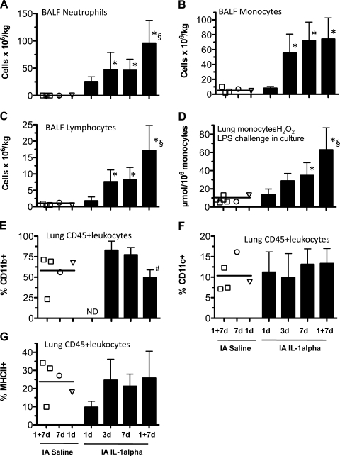
Exposure to IL-1α did not change body weight or lung weight-to-body weight ratio (Table 1). Intra-amniotic IL-1α did not increase BALF protein (the 2-fold increase at 3 days was not statistically significant), suggesting minimal lung injury. No neutrophils and very few monocytes or lymphocytes were detected in the BALF of control lambs (Fig. 1, A–C). IL-1α recruited neutrophils, monocytes, and lymphocytes to the air spaces by 3 days. Interestingly, the neutrophil and lymphocyte counts further increased twofold after repeated exposure to intra-amniotic IL-1α 7 days and again 1 day before delivery, but monocyte numbers did not change. The ability of lung monocytes to respond to LPS in vitro was evaluated by measurement of oxidant responses. While the immature lung monocytes from control lambs secreted 8 ± 1.4 μmol H2O2/106 monocytes, intra-amniotic IL-1α exposure for 7 days increased H2O2 secretion three- to fourfold, indicating maturation of the cells (Fig. 1D). Consistent with cell recruitment to BALF, repeated exposure to IL-1α further increased H2O2 generation twofold.
Fig. 1.
Intra-amniotic IL-1α-induced lung inflammation. Pregnant sheep were given intra-amniotic injections of recombinant sheep IL-1α 1, 3, or 7 days and repeat injections 7 days and again 1 day before delivery (1d, 3d, 7d, and 1+7d) prior to delivery at 124 ± 1 days. Controls received intra-amniotic saline (see Fig. 2 legend for symbols). A–C: bronchoalveolar lavage fluid (BALF) cells (neutrophils, monocytes, and lymphocytes) from fetal lungs. D: H2O2 generation in monocytes from fetal lung. Monocytes were purified over Percoll gradients and then challenged in culture with 100 ng/ml LPS, and H2O2 generation was measured and normalized to number of cells in culture. E–G: immunophenotyping for CD11b, CD11c, and major histocompatability complex class II (MHCII) in lung leukocytes purified using a CD45-biotin magnetically coupled antibody after enzymatic digestion. *P < 0.05 vs. composite control group. §P < 0.05 vs. IL-1α 1d. #P < 0.05 vs. IL-1α 7d. ND, not done; IA, intra-amniotic.
To better define the leukocyte populations, flow cytometry was performed to immunophenotype cells not recovered in the BALF by elastase digestion of lung tissue and purification of leukocytes using CD45 magnetic beads. CD11b, an integrin that mediates leukocyte adhesion and migration, is expressed on monocytes and activated neutrophils (41). Compared with controls, expression of CD11b (Fig. 1E), CD11c (Fig. 1F), or MHCII (Fig. 1G) did not change significantly after IL-1α exposure.
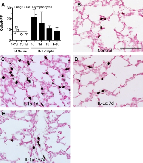
Next, we performed immunostaining with an antibody against the T-cell receptor CD3, which demonstrated recruitment of T lymphocytes 1 day after exposure, with no further increases after repeated IL-1α exposures (Fig. 2). Most of the CD3-positive T lymphocytes were in the pulmonary interstitium. Expression of MPO in neutrophils and monocytes also increased after IL-1α exposure (data not shown).
Fig. 2.
Intra-amniotic IL-1α recruited T lymphocytes to the lung. Pregnant sheep were given 1 or 2 intra-amniotic injections of recombinant sheep IL-1α for intervals shown prior to delivery at 124 ± 1 days; controls received intra-amniotic saline [1 + 7 days (□), 7 days (○), 1 day (▿)] prior to delivery at 124 ± 1 days. Lung sections from the fetuses were immunostained with an anti-CD3 antibody. A: quantitation of CD3-positive cells per high-power field (HPF). *P < 0.05 vs. composite control group. B–E: representative images from controls and fetuses exposed to IL-1α for 1 day, 7 days, and 1 + 7 days. Scale bar, 50 μm.
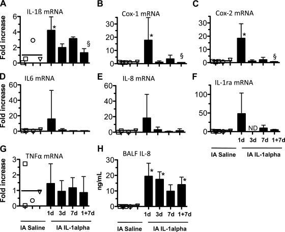
Intra-amniotic injection of IL-1α induced a threefold increase in expression of IL-1β and a 20-fold increase in the cyclooxygenase (COX) enzymes COX-1 and COX-2 in the lung 1 day after exposure (Fig. 3, A–C). IL-6, IL-8, and IL-1R antagonist mRNA expression in the lung increased variably 1 day after IL-1α, but these values were not significantly increased over controls (P=0.2, 0.07, and 0.3 vs. control, respectively; Fig. 3, D–F). IL-1α exposure also did not induce TNFα (Fig. 3G) or mRNA expression of the interferon-inducible genes IP-10 (CXCL10) and MIG (CXCL9) in the lung (data not shown). In contrast to the single exposure, cytokine mRNA expression after repeated exposure to intra-amniotic IL-1α was not different from controls. IL-8 protein was not detectable in the BALF of the control lambs, while IL-8 was present at 10–20 ng/ml in the BALF of all the groups subjected to single and repeated exposures to intra-amniotic IL-1α (Fig. 3H).
Fig. 3.
Intra-amniotic IL-1α increased pulmonary cytokine expression. Pregnant sheep were given 1 or 2 intra-amniotic injections of recombinant sheep IL-1α for intervals shown prior to delivery at 124 ± 1 days; controls received intra-amniotic saline (see symbols in Fig. 2 legend). A–G: after delivery, total RNA was extracted from fetal lung, and cytokines were quantified using real-time PCR assays with sheep-specific primers and TaqMan probes [IL-1β, cyclooxygenase enzymes (COX-1 and COX-2), IL-6, IL-8, IL-1 receptor antagonist (IL-1RA), and TNFα]. Values for each cytokine were normalized to 18S rRNA. Mean mRNA signals in control animals were given the value of 1, and levels at each time point are expressed relative to controls. H: BALF was used for IL-8 protein measurement using ELISA. *P < 0.05 vs. composite control group. §P < 0.05 vs. IL-1 1d. ND, not done.
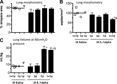
Lung morphometry was analyzed on the right upper lobe. Compared with controls, the air space fraction decreased (Fig. 4A) and the tissue fraction increased (data not shown) 1 day after intra-amniotic IL-1α. However, the air space fraction returned to control values at 3 and 7 days after IL-1α and after repeated exposures to IL-1α 7 days and again 1 day before delivery. Secondary alveolar septal crest density is a sensitive measure of lung development during the alveolar stage of lung development (4). Compared with controls, secondary alveolar septal crest density relative to air space area did not change significantly after a single exposure to intra-amniotic IL-1α (Fig. 4B; secondary septal crest density tended to be lower 3 days after IL-1α exposure, P=0.07 vs. controls). However, repeated exposure to intra-amniotic IL-1α 7 days and again 1 day before delivery decreased secondary septal crest density by 33% compared with controls. Lung gas volume increased 7 days after intra-amniotic IL-1α exposure. Lung volumes were also increased after repeated exposure to IL-1α 7 days and again 1 day before delivery, similar to a single exposure 7 days before delivery (Fig. 4C).
Fig. 4.
Intra-amniotic IL-1α increased lung volumes with modest effects on morphology. Pregnant sheep were given 1 or 2 intra-amniotic injections of recombinant sheep IL-1α for intervals shown prior to delivery at 124 ± 1 days; controls received intra-amniotic saline (see symbols in Fig. 2 legend). Right upper lobe of the lung was inflation-fixed at 30 cmH2O pressure for morphology. A: air space fraction expressed relative to total lung area. B: alveolar secondary septal crest density expressed relative to air space area. C: lung air volumes measured at 40 cmH2O pressure. *P < 0.05 vs. composite control group. §P < 0.05 vs. IL-1α 1d. #P < 0.05 vs. IL-1α 7d.
Systemic inflammatory responses after intra-amniotic IL-1α.
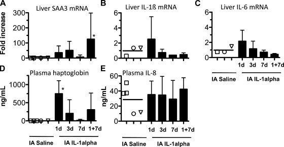
mRNA expression of the acute-phase reactant SAA tended to increase 35- to 50-fold in the liver 1 and 3 days (P=not significant) after IL-1α exposure (Fig. 5A). However, in the fetal lambs exposed to IL-1α 7 days and again 1 day before delivery, SAA expression in the liver increased. Another acute-phase reactant, plasma haptoglobin, increased from nearly undetectable levels in controls to 750 ± 135 ng/ml at 1 day, with a decrease to near control levels by 7 days (Fig. 5D). In the lambs exposed to IL-1α 7 days and again 1 day before delivery, plasma haptoglobin levels were 308 ± 460 ng/ml. Intra-amniotic IL-1α did not induce hepatic expression of IL-1β or IL-6 mRNA or increase plasma IL-8 protein levels (Fig. 5, B, C, and E). IL-1α also did not increase expression of TNFα, IFNγ, IL-4, IL-10, or IL-17 mRNA in the fetal spleen (data not shown). Intra-amniotic IL-1α tended to cause an initial neutropenia (3-fold decrease at 1 day, P=not significant; Table 2). Compared with controls, repeated exposure to IL-1α caused a 10-fold increase in neutrophil counts. Compared with controls, the monocyte counts increased sixfold 3 days after IL-1α exposure, but expression of CD14, a LPS coreceptor, lymphocyte counts, or the CD4-to-CD8 ratio did not change significantly after IL-1α exposure.
Fig. 5.
Intra-amniotic IL-1α induced modest systemic fetal inflammation. Pregnant sheep were given 1 or 2 intra-amniotic injections of recombinant sheep IL-1α for intervals shown prior to delivery at 124 ± 1 days; controls received intra-amniotic saline (see symbols in Fig. 2 legend). A: serum amyloid A3 (SAA3) in fetal liver was measured using an RNase protection assay. Values were normalized to L32, a ribosomal protein mRNA. B and C: IL-1β and IL-6 mRNA expression was measured using real-time PCR assays and TaqMan probes. Values were normalized to 18S rRNA. Mean mRNA signal in control animals was given the value of 1, and levels at each time point are expressed relative to controls. All assays used sheep-specific probes. D and E: plasma levels of haptoglobin and IL-8. *P < 0.05 vs. composite control group.
Table 2.
Changes in blood leukocytes and immunophenotype after intra-amniotic exposure to IL-1α
| IL-1α |
|||||
|---|---|---|---|---|---|
| Control Saline | 1 day | 3 days | 7 days | 7 days + 1 day | |
| Total WBC, 109/l | 6.4±6.3 | 3.9±0.7 | 7.1±3.3 | 4.4±1.7† | 18.6±16.4* |
| Neutrophils, 109/l | 1.4±1.4 | 0.4±0.4† | 3.1±1.7 | 2.3±1.0 | 14.9±14.8* |
| Lymphocytes, 109/l | 4.2±4.1 | 3.1±0.7 | 2.4±1.1 | 1.6±0.8 | 1.7±0.8 |
| Monocytes, 109/l | 0.2±0.07 | 0.1±0.08 | 1.3±0.8* | 0.1±0.09 | 0.1±0.08 |
| %CD14+ | 1.6±0.2 | 7.0±5.1† | 3.5±1.6 | 1.4±1.0 | 0.9±1.5 |
| %MHCII+ | 9.4±1.1 | 9±5 | 5±2 | ND | 22±16 |
| CD4-to-CD8 ratio | 1.5±0.2 | 1.1±0.5 | 1.8±0.3 | 0.8±0.2† | 1.7±0.6 |
Values are means ± SD. Control group is a composite of intra-amniotic saline 1 day + 7 days (n=3), 7 days (n=1), and 1 day (n=1). WBC, white blood cells; MCHII, major histocompatability complex class II; ND, not done.
P < 0.05 vs. control.
P < 0.05 vs. IL-1α (1 day + 7 days).
Inflammatory responses in the posterior mediastinal lymph node.
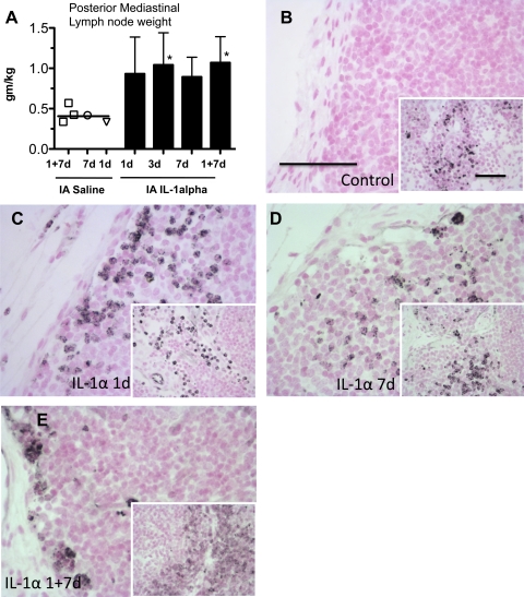
The posterior mediastinal lymph node receives afferent lymphatics from the lung and the gastrointestinal tract. In all the IL-1α-exposed groups (single as well as repeated exposures), lymph node weight doubled relative to the controls (Fig. 6A). iNOS (NOSII)-positive cells, most likely monocytes and neutrophils, were detected in the subcapsular and parafollicular regions of the lymph node in the IL-1α-exposed animals, but not controls (Fig. 6, B–E). In contrast, NOSII-positive cells were observed in the medullary regions of the lymph node in all groups. These results are consistent with migration of activated inflammatory cells from the lung or the gastrointestinal tract to this draining node (51). MPO-positive cells were increased in the subcapsular and paracortical regions after IL-1α exposure (data not shown). T-regulatory cells suppress inflammatory responses and are characterized by expression of the transcription factor Foxp3 (9). Foxp3-positive cells decreased by 75% at 1 and 3 days after intra-amniotic IL-1α, with return to baseline by 7 days (Table 3). In lambs exposed to IL-1α 7 days and again 1 day before delivery, numbers of Foxp3-positive cells in the posterior mediastinal lymph node were similar to controls.
Fig. 6.
Intra-amniotic IL-1α caused inflammation in posterior mediastinal lymph node. Pregnant sheep were given 1 or 2 intra-amniotic injections of recombinant sheep IL-1α for intervals shown prior to delivery at 124 ± 1 days; controls received intra-amniotic saline (see symbols in Fig. 2 legend). A: weight of posterior mediastinal lymph node normalized to body weight. B–E: representative images of immunostaining with an anti-inducible nitric oxide synthase (NOSII) antibody using lymph nodes from controls and animals exposed to IL-1α. Main frame of each panel shows subcapsular region; inset shows parafollicular and medullary areas. Scale bar, 50 μm. *P < 0.05 vs. composite control group. Note NOSII-positive cells only in IL-1α-exposed animals in subcapsular regions and medullary NOSII-positive cells in all groups.
Table 3.
Expression of Foxp3 in posterior mediastinal lymph node cells after intra-amniotic exposure to IL-1α
| Group | Foxp3+ Cells, %/HPF |
|---|---|
| Control saline | |
| 1 day + 7 days | 16.3, 20.7 |
| 7 days | 15.3 |
| 1 day | 18.6 |
| Composite | 17.8±2.3 |
| IL-1α | |
| 1 day | 3.6±3.6* |
| 3 days | 4.0±3.6* |
| 7 days | 12.6±3.0 |
| 1 day + 7 days | 19.4±6.4 |
Individual value are shown for control animals; other values are means ± SD. Immunohistology for Foxp3 was done using paraffin-embedded sections of posterior mediastinal lymph nodes. HPF, high-power field.
P < 0.05 vs. composite control group.
DISCUSSION
Inflammatory responses vary greatly on the basis of the route of exposure to the inflammatory agonists. For instance, IL-1 given intravenously (5–10 ng/kg) in humans elicits a pyrogenic response (8), and an intravascular injection of 10 μg of IL-1α in the sheep fetus is lethal (27), while a 10-fold-higher dose given by intra-amniotic injection in the present study did not result in lethality. We used recombinant sheep IL-1α for these experiments, but recombinant sheep IL-1β also results in similar lung inflammatory responses (54). In contrast to the intravenous injections, fetal inflammation resulting from agonists injected into the amniotic fluid is mediated via contact of the agents with the lung, chorioamnion, gastrointestinal, and skin epithelia (29, 33, 56). A biologically important conclusion from the present study is that intra-amniotic injection of IL-1α can induce a unique inflammatory response in which the pulmonary inflammation is robust, while systemic inflammatory responses are more modest. The inflammatory responses in fetal lambs are similar to that in human infants exposed to chorioamnionitis caused by microorganisms signaling via different TLRs (14, 42).
A striking result of our study is that, as a consequence of lung inflammation, lung volumes increased by about threefold 7 days after intra-amniotic IL-1α exposure, without significant changes in lung structure. Acute lung inflammation following IL-1α exposure induced increases in cytokine expression and recruitment of leukocytes, resulting in decreased air space fraction (and increased tissue fraction) 1 day after IL-1α exposure. However, the air space fraction normalized at later times after IL-1α exposure, presumably secondary to resolution of tissue edema. We previously reported increased surfactant pools 7 days after intra-amniotic IL-1α in the fetal sheep (49, 54). Taken together, the increased lung volumes after IL-1α are likely due to surfactant, rather than anatomic, effects. The fetal sheep at 124 days gestation is in an alveolar stage of lung development (1). Therefore, any perturbation in lung development should be reflected in arrested development of alveolar secondary septal crests. Interestingly, the secondary septal crest density did not decrease following a single exposure to IL-1α but did decrease by ∼33% following repeated exposures to IL-1α. We previously reported a more significant impairment of alveolar development after intra-amniotic LPS exposure in fetal sheep, with a similar increase in lung volume (53).
Unlike the adult, the preterm sheep fetus is able to respond to a limited number of proinflammatory stimuli injected into the amniotic fluid. While a TLR4 agonist, LPS, induced a robust inflammatory response, a TLR2 agonist caused an inconsistent inflammatory response and a TLR3 agonist did not cause fetal inflammation (16). Similarly, intra-amniotic injection of IL-8 or TNFα induced mild or no inflammation (18, 25). However, there appear to be some species differences: Sadowsky et al. (47) demonstrated that intra-amniotic infusions of IL-1β and TNFα, but not IL-6 or IL-8, induced preterm labor in nonhuman primates. The present study demonstrated that IL-1α is a unique proinflammatory cytokine, in that it can induce robust fetal inflammation after intra-amniotic injection.
The IL-1 cytokine family includes IL-1α, IL-1β, IL-1R antagonist, IL-18, IL-33, and others (6). IL-1α and IL-1β are synthesized as precursor molecules. While IL-1α is a cell-associated cytokine that is active as a precursor, IL-1β is active only after the precursor is cleaved and the cytokine is secreted. The recombinant sheep IL-1α used in this study was the mature protein and was used because previous dose-finding experiments demonstrated that IL-1α was more potent than IL-1β in this model (54). Since both cytokines activate the common IL-1R (6), the present study demonstrates that IL-1 signaling in the fetus can induce a systemic fetal inflammatory response.
There are similarities between the IL-1α- and LPS-induced fetal inflammatory responses. This is not surprising, because LPS receptor signaling via TLR4 and IL-1 signaling via IL-1R share similar intracellular signaling pathways (38). Similar to the previously reported responses to intra-amniotic LPS (28, 34), we demonstrate here that intra-amniotic IL-1α induced an equivalent recruitment of neutrophils, monocytes, and T lymphocytes to the lung in sheep fetuses. These inflammatory cells were activated because they expressed NOSII and MPO. IL-1α exposure induced the cyclooxygenase enzymes Cox-1 and Cox-2 and caused the lung monocytes to mature. The maturation of lung monocytes was demonstrated by a minimal oxidative burst in the control monocytes, while IL-1α exposure in vivo for 3 or 7 days increased H2O2 secretion by the lung monocytes in response to an in vitro challenge with LPS. The IL-1α-induced lung inflammation resulted in increased lung gas volumes, similar to LPS effects (28, 34). However, unlike LPS, intra-amniotic IL-1α induction of IL-1β, IL-6, and IL-8 mRNA in the lung was minimal. The reasons for the decreased induction of proinflammatory cytokine expression by IL-1α are not known. Lack of protein data for many cytokines may obscure interpretation of inflammatory effects of intra-amniotic IL-1α. LPS induces activation of the transcription factor NF-κB via MyD88-dependent and Toll/IL-1 receptor domain-containing adaptor-inducing IFNβ (TRIF)-mediated MyD88-independent pathways (38). The TRIF-mediated NF-κB activation induces interferon, which increases the expression of interferon-inducible chemokines such as IP-10 and MIG. We previously demonstrated that intra-amniotic LPS induces IP-10 and MIG in the fetal sheep lung (23), while no induction of these interferon-inducible chemokines after IL-1α exposure was observed in this study. Taken together, these results demonstrate that although signaling of LPS and IL-1 in the fetus is similar, some aspects of their inflammatory repertoire are unique.
Fetal inflammatory response syndrome (FIRS) in the human is a unique systemic inflammatory response defined as chorioamnionitis associated with cord plasma IL-6 levels >11 pg/ml (14, 45). Unlike the “cytokine storm,” which causes multiorgan dysfunction associated with the systemic inflammatory response syndrome in adults (40), FIRS is a more subtle inflammatory response. Despite the modest inflammatory responses, FIRS was postulated to be the proximate cause of multiple adverse neonatal outcomes (15). Since <2% of the preterm infants exposed to chorioamnionitis have early-onset bacteremia (50), systemic inflammation in the fetus must be initiated by inflammatory responses in the fetal organs in contact with infected amniotic fluid. Indeed, we and others have demonstrated that the fetal chorioamnion, lung, gut, and skin contribute to the systemic inflammation induced by chorioamnionitis (30, 33). Chorioamnionitis in humans is caused by a variety of organisms, mostly of low-grade virulence that colonize the female lower genital tract (42).
In the present study, IL-1α induced a systemic inflammatory response. Compared with the pulmonary responses, the systemic inflammation was modest. The neutrophil counts in the blood changed, and the expression of the acute-phase reactant haptoglobin in plasma increased. Interestingly, while the total lymphocyte counts in the blood did not change, the CD4-to-CD8 ratio appeared to decrease in the cord blood 7 days after IL-1α exposure. A decrease in the CD4-to-CD8 ratio in the peripheral blood can be induced by a number of chronic infections (46). One caveat of interpretation of data from FACSCanto analysis is that the blood leukocyte counts initially decreased and subsequently increased. Therefore, some of the reported change in expression of leukocyte markers could be confounded by this changing fetal blood leukocyte count. One question regarding systemic inflammation relates to its pathogenesis. Using fetal surgical techniques, we recently selectively infused LPS into the fetal airways or the amniotic fluid, with occlusion of the airway to prevent LPS from contacting the lung (33). Lung infusion and chorioamnion exposure to LPS can induce a systemic fetal inflammatory response. These experiments illustrate the complex signaling between different fetal organs, resulting in fetal inflammation from chorioamnionitis.
IL-1α also caused inflammatory changes in the lymph node that drains lymph from the lung and the gastrointestinal tract. After IL-1α exposure, the weight of the lymph node doubled, and NOSII- and MPO-staining cells were detected in the subcapsular space and the parafollicular and medullary regions. These results suggest migration of activated inflammatory cells from the draining organs and activation of the resident macrophages. Similar to an intra-amniotic LPS-induced reduction of Foxp3-positive T-regulatory lymphocytes in the fetal sheep thymus (35), we demonstrated a time-dependent decrease in Foxp3-staining cells in the posterior mediastinal lymph node. Foxp3 is a master regulator of the anti-inflammatory T-regulatory cells (10, 17). However, a detailed characterization of T-regulatory cells was not possible because of lack of appropriate cross-reactive antibodies for sheep. Although the findings are preliminary, the experiment suggests that IL-1α signaling in chorioamnionitis can induce a proinflammatory fetal milieu, since T-regulatory lymphocytes are known to dampen inflammatory responses (9). Despite the changes in the blood, liver, and regional lymph node, we did not observe significant increases in the Th1, Th2, or Th17 cytokines in the spleen after exposure to intra-amniotic IL-1α. Because of limitations of reagents for sheep dendritic cells, we could not determine if there was migration and signaling via dendritic cells. Whether the inflamed lymph node mediates systemic inflammation or whether the changes in the lymph node are merely a response to end-organ inflammation is unknown.
The duration of exposure to chorioamnionitis in humans is often prolonged (43); therefore, exposure to proinflammatory agonists can also be for extended periods. We previously demonstrated that repeated exposures to LPS induced tolerance to endotoxin and other TLR agonists (22, 32). We therefore asked if repeated exposure to IL-1α would cause innate immune tolerance. Repeated injections of IL-1α decreased some measurements of inflammatory response but not others. Repeated exposure to intra-amniotic IL-1α increased inflammatory cell recruitment into the fetal lung, induced the monocyte oxidant response, and increased blood neutrophils, suggesting no tolerance. However, unlike the single IL-1α exposure, repeated exposure to IL-1α did not increase lung COX expression or decrease T-regulatory lymphocytes in the posterior mediastinal lymph node. Therefore, unlike our previous results with LPS (22), this study demonstrated complex organ- and pathway-specific effects after repeated exposure to IL-1α. The present study demonstrates that IL-1α, a cytokine that can be induced by multiple different pathogens, is a mediator of the FIRS.
GRANTS
This study was funded by National Institute of Child Health and Human Development Grant HD-57869 (to S. G. Kallapur).
DISCLOSURES
No conflicts of interest, financial or otherwise, are declared by the authors.
ACKNOWLEDGMENTS
The authors thank Dr. Kathy Heel and Tracey Lee-Pullen (Center for Microscopy Characterization and Analysis, University of Western Australia) for consultation on FACS experiments; Dr. Susan Wert for consultation on morphometry and Dr. Jareen Meinzen-Derr for statistics consultation (University of Cincinnati); and Amy Whitescarver, Manuel Alvarez, Jr., and Avedis Kazanjian for expert technical assistance.
REFERENCES
- 1. Alcorn DG, Adamson TM, Maloney JE, Robinson PM. A morphologic and morphometric analysis of fetal lung development in the sheep. Anat Rec 201: 655–667, 1981 [DOI] [PubMed] [Google Scholar]
- 2. Berry CA, Nitsos I, Hillman NH, Pillow JJ, Polglase GR, Kramer BW, Kemp MW, Newnham JP, Jobe AH, Kallapur SG. Interleukin 1 in lipopolysaccharide induced chorioamnionitis in the fetal sheep. Reprod Sci. In press [DOI] [PMC free article] [PubMed] [Google Scholar]
- 3. Bry K, Whitsett JA, Lappalainen U. IL-1β disrupts postnatal lung morphogenesis in the mouse. Am J Respir Cell Mol Biol 36: 32–42, 2007 [DOI] [PMC free article] [PubMed] [Google Scholar]
- 4. Burri PH. Fetal and postnatal development of the lung. Annu Rev Physiol 46: 617–628, 1984 [DOI] [PubMed] [Google Scholar]
- 5. Chen GY, Nunez G. Sterile inflammation: sensing and reacting to damage. Nat Rev Immunol 10: 826–837, 2010 [DOI] [PMC free article] [PubMed] [Google Scholar]
- 6. Dinarello CA. Immunological and inflammatory functions of the interleukin-1 family. Annu Rev Immunol 27: 519–550, 2009 [DOI] [PubMed] [Google Scholar]
- 7. Dinarello CA. Interleukin-1β and the autoinflammatory diseases. N Engl J Med 360: 2467–2470, 2009 [DOI] [PubMed] [Google Scholar]
- 8. Dinarello CA, Renfer L, Wolff SM. Human leukocytic pyrogen: purification and development of a radioimmunoassay. Proc Natl Acad Sci USA 74: 4624–4627, 1977 [DOI] [PMC free article] [PubMed] [Google Scholar]
- 9. Fehervari Z, Sakaguchi S. CD4+ T-regs and immune control. J Clin Invest 114: 1209–1217, 2004 [DOI] [PMC free article] [PubMed] [Google Scholar]
- 10. Fontenot JD, Gavin MA, Rudensky AY. Foxp3 programs the development and function of CD4+CD25+ regulatory T cells. Nat Immunol 4: 330–336, 2003 [DOI] [PubMed] [Google Scholar]
- 11. Gasse P, Mary C, Guenon I, Noulin N, Charron S, Schnyder-Candrian S, Schnyder B, Akira S, Quesniaux VF, Lagente V, Ryffel B, Couillin I. IL-1R1/MyD88 signaling and the inflammasome are essential in pulmonary inflammation and fibrosis in mice. J Clin Invest 117: 3786–3799, 2007 [DOI] [PMC free article] [PubMed] [Google Scholar]
- 12. Genc MR, Gerber S, Nesin M, Witkin SS. Polymorphism in the interleukin-1 gene complex and spontaneous preterm delivery. Am J Obstet Gynecol 187: 157–163, 2002 [DOI] [PubMed] [Google Scholar]
- 13. Goldenberg RL, Hauth JC, Andrews WW. Intrauterine infection and preterm delivery. N Engl J Med 342: 1500–1507, 2000 [DOI] [PubMed] [Google Scholar]
- 14. Gomez R, Romero R, Ghezzi F, Yoon B, Mazor M, Berry S. The fetal inflammatory response syndrome. Am J Obstet Gynecol 179: 194–202, 1998 [DOI] [PubMed] [Google Scholar]
- 15. Gotsch F, Romero R, Kusanovic JP, Mazaki-Tovi S, Pineles BL, Erez O, Espinoza J, Hassan SS. The fetal inflammatory response syndrome. Clin Obstet Gynecol 50: 652–683, 2007 [DOI] [PubMed] [Google Scholar]
- 16. Hillman NH, Moss TJ, Nitsos I, Kramer BW, Bachurski CJ, Ikegami M, Jobe AH, Kallapur SG. Toll-like receptors and agonist responses in the developing fetal sheep lung. Pediatr Res 63: 388–393, 2008 [DOI] [PubMed] [Google Scholar]
- 17. Hori S, Nomura T, Sakaguchi S. Control of regulatory T cell development by the transcription factor Foxp3. Science 299: 1057–1061, 2003 [DOI] [PubMed] [Google Scholar]
- 18. Ikegami M, Moss TJ, Kallapur SG, Mulrooney N, Kramer BW, Nitsos I, Bachursky CJ, Newnham JP, Jobe AH. Minimal lung and systemic responses to TNFα in preterm sheep. Am J Physiol Lung Cell Mol Physiol 285: L121–L129, 2003 [DOI] [PubMed] [Google Scholar]
- 19. Jobe AH, Newnham JP, Willet KE, Moss TJ, Ervin MG, Padbury JF, Sly PD, Ikegami M. Endotoxin-induced lung maturation in preterm lambs is not mediated by cortisol. Am J Respir Crit Care Med 162: 1656–1661, 2000 [DOI] [PubMed] [Google Scholar]
- 20. Kakkera DK, Siddiq MM, Parton LA. Interleukin-1 balance in the lungs of preterm infants who develop bronchopulmonary dysplasia. Biol Neonate 87: 82–90, 2005 [DOI] [PubMed] [Google Scholar]
- 21. Kallapur SG, Ikegami M. Physiological consequences of intrauterine insults. Paediatr Respir Rev 7: 110–116, 2006 [DOI] [PubMed] [Google Scholar]
- 22. Kallapur SG, Jobe AH, Ball MK, Nitsos I, Moss TJ, Hillman NH, Newnham JP, Kramer BW. Pulmonary and systemic endotoxin tolerance in preterm fetal sheep exposed to chorioamnionitis. J Immunol 179: 8491–8499, 2007 [DOI] [PubMed] [Google Scholar]
- 23. Kallapur SG, Jobe AH, Ikegami M, Bachurski CJ. Increased IP-10 and MIG expression after intra-amniotic endotoxin in preterm lamb lung. Am J Respir Crit Care Med 167: 779–786, 2003 [DOI] [PubMed] [Google Scholar]
- 24. Kallapur SG, Kramer BW, Moss TJ, Newnham JP, Jobe AH, Ikegami M, Bachurski CJ. Maternal glucocorticoids increase endotoxin-induced lung inflammation in preterm lambs. Am J Physiol Lung Cell Mol Physiol 284: L633–L642, 2003 [DOI] [PubMed] [Google Scholar]
- 25. Kallapur SG, Moss TJ, Auten RL, Jr, Nitsos I, Pillow JJ, Kramer BW, Maeda DY, Newnham JP, Ikegami M, Jobe AH. IL-8 signaling does not mediate intra-amniotic LPS-induced inflammation and maturation in preterm fetal lamb lung. Am J Physiol Lung Cell Mol Physiol 297: L512–L519, 2009 [DOI] [PMC free article] [PubMed] [Google Scholar]
- 26. Kallapur SG, Moss TJM, Ikegami M, Jasman RL, Newnham JP, Jobe AH. Recruited inflammatory cells mediate endotoxin induced lung maturation in preterm fetal lambs. Am J Respir Crit Care Med 172: 1315–1321, 2005 [DOI] [PMC free article] [PubMed] [Google Scholar]
- 27. Kallapur SG, Nitsos I, Moss TJ, Polglase GR, Pillow JJ, Cheah FC, Kramer BW, Newnham JP, Ikegami M, Jobe AH. IL-1 mediates pulmonary and systemic inflammatory responses to chorioamnionitis induced by lipopolysaccharide. Am J Respir Crit Care Med 179: 955–961, 2009 [DOI] [PMC free article] [PubMed] [Google Scholar]
- 28. Kallapur SG, Willet KE, Jobe AH, Ikegami M, Bachurski CJ. Intra-amniotic endotoxin: chorioamnionitis precedes lung maturation in preterm lambs. Am J Physiol Lung Cell Mol Physiol 280: L527–L536, 2001 [DOI] [PubMed] [Google Scholar]
- 29. Kemp MW, Saito M, Nitsos I, Jobe AH, Kallapur S, Newnham JP. Exposure to in utero lipopolysaccharide induces inflammation in the fetal ovine skin. Reprod Sci 18: 88–98, 2011 [DOI] [PMC free article] [PubMed] [Google Scholar]
- 30. Kim YM, Romero R, Chaiworapongsa T, Espinoza J, Mor G, Kim CJ. Dermatitis as a component of the fetal inflammatory response syndrome is associated with activation of Toll-like receptors in epidermal keratinocytes. Histopathology 49: 506–514, 2006 [DOI] [PMC free article] [PubMed] [Google Scholar]
- 31. Kramer BW, Joshi SN, Moss TJM, Newnham JP, Sindelaar R, Jobe AH, Kallapur SG. Endotoxin-induced maturation of monocytes in preterm sheep lung. Am J Physiol Lung Cell Mol Physiol 293: L345–L353, 2007 [DOI] [PubMed] [Google Scholar]
- 32. Kramer BW, Kallapur SG, Moss TJ, Nitsos I, Newnham JP, Jobe AH. Intra-amniotic LPS modulation of TLR signaling in lung and blood monocytes of fetal sheep. Innate Immun 15: 101–107, 2009 [DOI] [PubMed] [Google Scholar]
- 33. Kramer BW, Kallapur SG, Moss TJ, Nitsos I, Polglase GP, Newnham JP, Jobe AH. Modulation of fetal inflammatory response on exposure to lipopolysaccharide by chorioamnion, lung, or gut in sheep. Am J Obstet Gynecol 202: e71–e79, 2010 [DOI] [PMC free article] [PubMed] [Google Scholar]
- 34. Kramer BW, Moss TJ, Willet KE, Newnham JP, Sly PD, Kallapur SG, Ikegami M, Jobe AH. Dose and time response for inflammation and lung maturation after intra-amniotic endotoxin in preterm lambs. Am J Respir Crit Care Med 164: 982–988, 2001 [DOI] [PubMed] [Google Scholar]
- 35. Kunzmann S, Glogger K, Been JV, Kallapur SG, Nitsos I, Moss TJ, Speer CP, Newnham JP, Jobe AH, Kramer BW. Thymic changes after chorioamnionitis induced by intraamniotic lipopolysaccharide in fetal sheep. Am J Obstet Gynecol 202: e471–e479, 2010 [DOI] [PMC free article] [PubMed] [Google Scholar]
- 36. Larsen CM, Faulenbach M, Vaag A, Volund A, Ehses JA, Seifert B, Mandrup-Poulsen T, Donath MY. Interleukin-1-receptor antagonist in type 2 diabetes mellitus. N Engl J Med 356: 1517–1526, 2007 [DOI] [PubMed] [Google Scholar]
- 37. Lee AJ, Lambermont VA, Pillow JJ, Polglase GR, Nitsos I, Newnham JP, Beilharz MW, Kallapur SG, Jobe AH, Kramer BW. Fetal responses to lipopolysaccharide-induced chorioamnionitis alter immune and airway responses in 7-week-old sheep. Am J Obstet Gynecol 204: 364. e317–e324, 2011 [DOI] [PubMed] [Google Scholar]
- 38. Li X, Qin J. Modulation of Toll-interleukin 1 receptor mediated signaling. J Mol Med 83: 258–266, 2005 [DOI] [PubMed] [Google Scholar]
- 39. Martinon F, Mayor A, Tschopp J. The inflammasomes: guardians of the body. Annu Rev Immunol 27: 229–265, 2009 [DOI] [PubMed] [Google Scholar]
- 40. Matsuda N, Hattori Y. Systemic inflammatory response syndrome (SIRS): molecular pathophysiology and gene therapy. J Pharm Sci 101: 189–198, 2006 [DOI] [PubMed] [Google Scholar]
- 41. Maus U, Huwe J, Ermert L, Ermert M, Seeger W, Lohmeyer J. Molecular pathways of monocyte emigration into the alveolar air space of intact mice. Am J Respir Crit Care Med 165: 95–100, 2002 [DOI] [PubMed] [Google Scholar]
- 42. Onderdonk AB, Delaney ML, DuBois AM, Allred EN, Leviton A. Detection of bacteria in placental tissues obtained from extremely low gestational age neonates. Am J Obstet Gynecol 198: e111–e117, 2008 [DOI] [PubMed] [Google Scholar]
- 43. Perni SC, Vardhana S, Korneeva I, Tuttle SL, Paraskevas LR, Chasen ST, Kalish RB, Witkin SS. Mycoplasma hominis and Ureaplasma urealyticum in midtrimester amniotic fluid: association with amniotic fluid cytokine levels and pregnancy outcome. Am J Obstet Gynecol 191: 1382–1386, 2004 [DOI] [PubMed] [Google Scholar]
- 44. Rindfleisch MS, Hasday JD, Taciak V, Broderick K, Viscardi RM. Potential role of interleukin-1 in the development of bronchopulmonary dysplasia. J Interferon Cytokine Res 16: 365–373, 1996 [DOI] [PubMed] [Google Scholar]
- 45. Romero R, Gomez R, Ghezzi F, Yoon B, Mazor M, Edwin S, Berry S. A fetal systemic inflammatory response is followed by the spontaneous onset of preterm parturition. Am J Obstet Gynecol 179: 186–193, 1998 [DOI] [PubMed] [Google Scholar]
- 46. Rosenberg YJ, Anderson AO, Pabst R. HIV-induced decline in blood CD4/CD8 ratios: viral killing or altered lymphocyte trafficking? Immunol Today 19: 10–17, 1998 [DOI] [PubMed] [Google Scholar]
- 47. Sadowsky DW, Adams KM, Gravett MG, Witkin SS, Novy MJ. Preterm labor is induced by intraamniotic infusions of interleukin-1β and tumor necrosis factor-α but not by interleukin-6 or interleukin-8 in a nonhuman primate model. Am J Obstet Gynecol 195: 1578–1589, 2006 [DOI] [PubMed] [Google Scholar]
- 48. Shah TA, Hillman NH, Nitsos I, Polglase GR, Pillow JJ, Newnham JP, Jobe AH, Kallapur SG. Pulmonary and systemic expression of monocyte chemotactic proteins in preterm sheep fetuses exposed to lipopolysaccharide-induced chorioamnionitis. Pediatr Res 68: 210–215, 2010 [DOI] [PMC free article] [PubMed] [Google Scholar]
- 49. Sosenko IR, Kallapur SG, Nitsos I, Moss TJ, Newnham JP, Ikegami M, Jobe AH. IL-1α causes lung inflammation and maturation by direct effects on preterm fetal lamb lungs. Pediatr Res 60: 294–298, 2006 [DOI] [PubMed] [Google Scholar]
- 50. Stoll BJ, Gordon T, Korones SB, Shankaran S, Tyson JE, Bauer CR, Fanaroff AA, Lemons JA, Donovan EF, Oh W, Stevenson DK, Ehrenkranz RA, Papile LA, Verter J, Wright LL. Early-onset sepsis in very low birth weight neonates: a report from the National Institute of Child Health and Human Development Neonatal Research Network. J Pediatr 129: 72–80, 1996 [DOI] [PubMed] [Google Scholar]
- 51. von Andrian UH, Mempel TR. Homing and cellular traffic in lymph nodes. Nat Rev Immunol 3: 867–878, 2003 [DOI] [PubMed] [Google Scholar]
- 52. Watterberg K, Demers L, Scott S, Murphy S. Chorioamnionitis and early lung inflammation in infants in whom bronchopulmonary dysplasia develops. Pediatrics 97: 210–215, 1996 [PubMed] [Google Scholar]
- 53. Willet KE, Jobe AH, Ikegami M, Brennan S, Newnham J, Sly PD. Antenatal endotoxin and glucocorticoid effects on lung morphometry in preterm lambs. Pediatr Res 48: 782–788, 2000 [DOI] [PubMed] [Google Scholar]
- 54. Willet KE, Kramer BW, Kallapur SG, Ikegami M, Newnham JP, Moss TJ, Sly PD, Jobe AH. Intra-amniotic injection of IL-1 induces inflammation and maturation in fetal sheep lung. Am J Physiol Lung Cell Mol Physiol 282: L411–L420, 2002 [DOI] [PubMed] [Google Scholar]
- 55. Wilson TC, Bachurski CJ, Ikegami M, Jobe AH, Kallapur SG. Pulmonary and systemic induction of SAA3 after ventilation and endotoxin in preterm lambs. Pediatr Res 58: 1204–1209, 2005 [DOI] [PubMed] [Google Scholar]
- 56. Wolfs TG, Buurman WA, Zoer B, Moonen RM, Derikx JP, Thuijls G, Villamor E, Gantert M, Garnier Y, Zimmermann LJ, Kramer BW. Endotoxin induced chorioamnionitis prevents intestinal development during gestation in fetal sheep. PLoS One 4: e5837, 2009 [DOI] [PMC free article] [PubMed] [Google Scholar]
- 57. Wolfs TG, Kallapur SG, Polglase GR, Pillow JJ, Nitsos I, Newnham JP, Chougnet CA, Kroon E, Spierings J, Willems CH, Jobe AH, Kramer BW. IL-1α mediated chorioamnionitis induces depletion of FoxP3+ cells and ileal inflammation in the ovine fetal gut. PLoS One 6: e18355, 2011 [DOI] [PMC free article] [PubMed] [Google Scholar]


