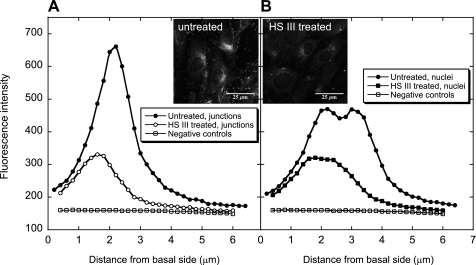
Fig. 6.
Confocal vertical profiles of HS before and after enzyme digestion with HS III (15 mU/ml) (n = 3). A: cell junction location. B: nuclear location. Negative controls were not treated with the enzyme or anti-HS, but only stained with the secondary anti-IgM-k antibody. The insets show confocal images of untreated and HS III-treated BLMVECs.

