Fig. 7.
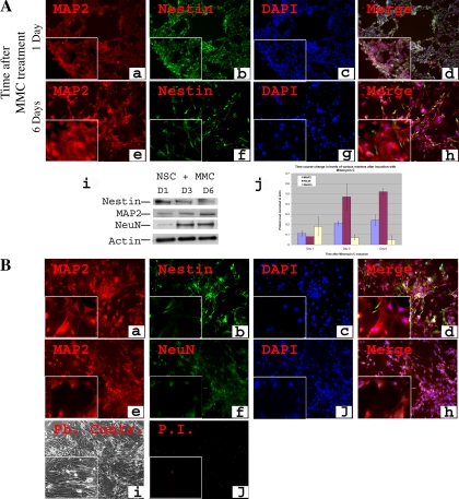
Generation and characterization of neuronal cells after MMC induction of NSCs. C17.2 NSCs were treated with 0.4 μg/ml of MMC under normoxic conditions, and immunostaining for the stem cell marker nestin and early neuronal marker NeuN is shown at 2 time points: 1 day and 6 days after treatment (A). Nestin was detected at day 1 (A, b) and decreased in intensity afterwards but remained detectable (A, f). MAP2 was weakly expressed at day 1 (A, a) and increased in intensity afterwards (A, e). Western blot analysis showed 2-fold increase in MAP2 from day 1 to day 3 and a significant (P < 0.005) ∼5-fold increase of NeuN from day 1 to day 6 (A, i, j). After 6 days of culture when the phenotype stabilized, cells were further cultured for an additional 2 days (total 8 days) and then characterized similarly. They were stained for nestin (B, b), MAP2 (B, a, e), and NeuN (B, f). Phase contrast microscopy confirmed the neuronal morphology (B, i). Mortality was only at a baseline level (B, j). Magnification: ×100; insets, ×400. Student's t-test, NeuN level at day 1 vs. day 6, P < 0.005.

