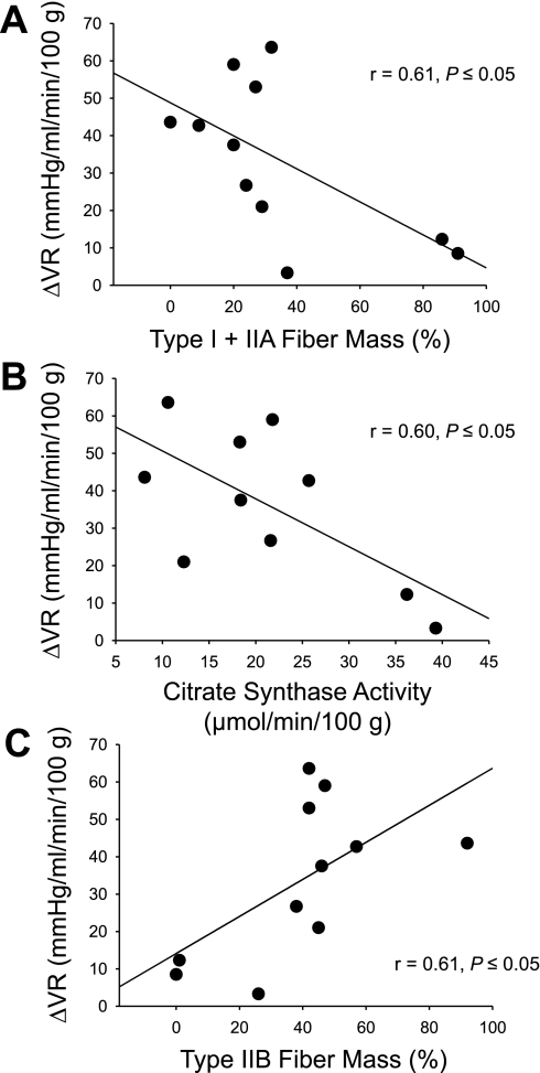
Fig. 3.
Relationships between the % sum of type I and IIA fibers (A), citrate synthase activity (B), and % sum of type IIB fibers (C) of the individual muscles listed in Table 1 of the rat hindlimb and the change in vascular resistance (ΔVR) after infusion of phenylephrine. Based on fiber-type composition and citrate synthase activity [reported by Delp and Duan (10)].

