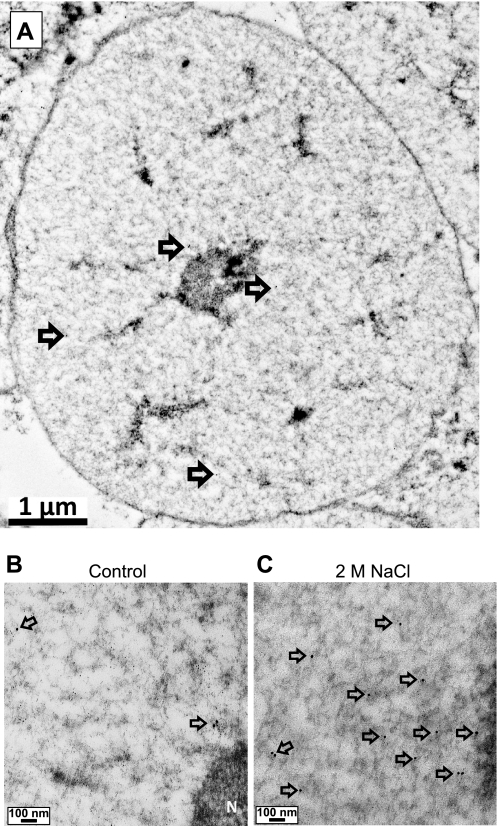
Fig. 1.
Low-power micrographs of isolated nuclei. A: illustration of a cell nucleus taken at low (6,000×) power to illustrate that intact nuclei were captured using the procedure. The nuclear membrane, nucleolus, and several 15-nm gold beads (arrows) may be detected. At 40,000×, a few NK3R 15-nm gold beads (arrows) are seen in the nucleus isolated from a control rat (B) and multiple NK3R 15 nm gold beads (arrows) are seen in the nucleus from a rat treated with 2 M NaCl (C). At these powers, however, 6-nm beads are difficult to discern.

