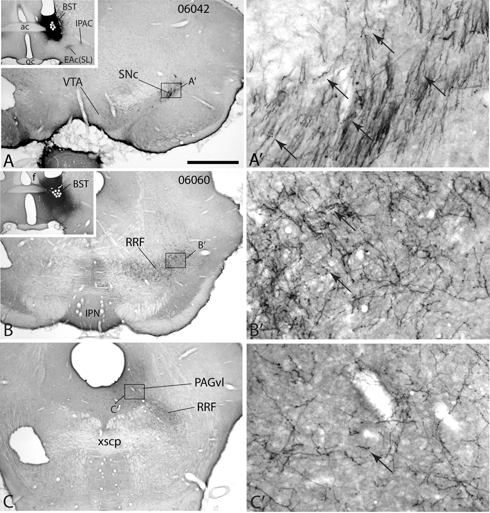
Figure 5.
Micrographs illustrating the distribution of anterogradely labeled axons in the midbrain dopaminergic complex in case 06042 (A-A’) in which an injection of PHA-L (black injection site) was placed in the lateral division of the bed nucleus of stria terminalis (BST, inset in A) and case 06060 (B–C’) with a PHA-L injection in the lateral division of the BST at a slightly more ventral position. White dots indicate the positions of PHA-L impregnated neurons marking the injection sites. A’, B’ and C’ are enlargements of the respective boxes in A, B and C. In case 06042 a rostral section through the midbrain dopaminergic complex (A and A’) exhibits dense anterograde labeling in the substantia nigra compacta (SNc) mostly in the form of non-varicose presumably passing fibers, although some varicosities (puncta) are visible (arrows). Little anterograde labeling is visible in the ventral tegmental area (VTA) in this case at this level. In case 06060, anterograde labeling spreads homogeneously through the retrorubral field (RRF, B and B’) and is more varicose (a few examples are arrowed). The ventrolateral periaqueductal gray substance (PAGvl) also contains a moderately dense plexus of varicose labeled fibers (C and C’). Additional abbreviations: ac – anterior commissure, EAc(SL) – central division of extended amygdala, sublenticular division; f – fornix; IPAC – interstitial nucleus of the posterior limb of the anterior commissure; IPN – interpeduncular nucleus; xscp – crossing of the superior cerebellar peduncle. Scale bar: 1 mm in A, B and C; 100 µm in A’, B’ and C’.

