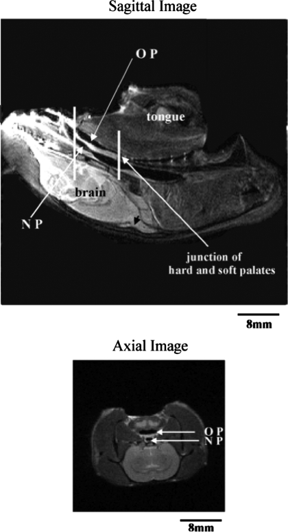
Fig. 1.
Top: sagittal MRI of the rat head, with tongue and brain labeled for orientation. Oropharynx (OP), nasopharynx (NP), and junction of the hard and soft palate are also indicated. Vertical lines show 8-mm region where axial slices were taken. Bottom: representative axial slice through the caudal part of the 8-mm region of interest.

