Fig. 4.
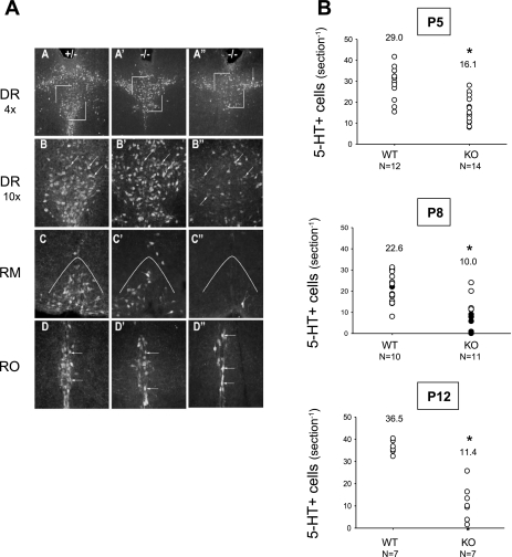
Pet-1−/− animals have reduced 5-HT-positive neurons in pons and medulla. 5-HT-positive cell bodies were quantified in the raphe magnus to represent 5-HT system status as a whole. A: 5-HT-positive neurons at P8 in the dorsal raphe (DR), raphe magnus (RM), and raphe obscurus (RO) of a wild-type littermate (A–D), a Pet-1−/− animal (−/−) surviving 4 episodes of anoxia (A'–D'), and a Pet-1−/− animal that did not survive anoxia (A''–D''). B: numbers of 5-HT-positive cell bodies in the raphe magnus of WT and Pet-1−/− animals (KO) at P5 (top), P8 (middle), and P12 (bottom). At P8, KO animals that die (●) have significantly fewer neurons than KO animals surviving (○). At all ages, KO animals have fewer 5-HT-positive neurons than WT (*P < 0.01, KO vs. WT) and at P8, KO animals dying have ∼1/3 the 5-HT neurons as those surviving. In B, numbers above each set of points are the average cell counts.

