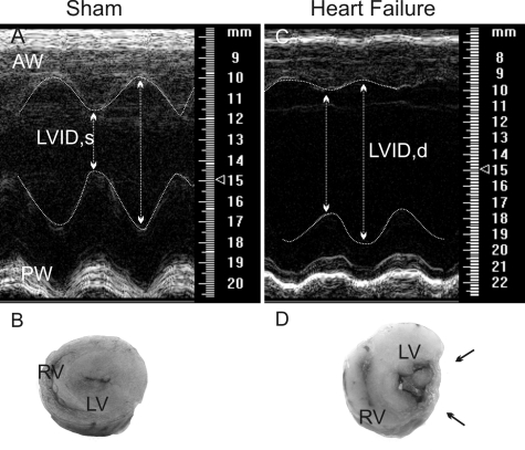
Fig. 1.
Echocardiographic assessment of left ventricular function. Representative echocardiograph images are movement-mode, parasternal short-axis views of a sham (A) and a heart failure (HF) rat (C). Macroscopic, transverse sections from representative hearts in a sham (B) and HF rat (D) are also shown. Note the thinning of the left ventricle infarcted wall (between arrows). AW, anterior wall; LVID,d and LVID,s: left ventricle internal dimension in diastole and systole; PW, posterior wall; LV, left ventricle; RV, right ventricle.

