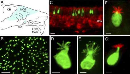
Fig. 1.
Identical properties shared by choline acetyltransferase green fluorescent protein (ChAT GFP)-expressing and transient receptor potential channel M5 (TRPM5)-expressing microvillous cells. A: schematic drawing of a mouse hemi-nose, showing distribution of GFP-expressing microvillous cells in the main olfactory epithelium (MOE) and septal organ (SO) (blue regions) in both ChAT(BAC)-enhanced GFP (eGFP) and TRPM5-GFP mice. OB, olfactory bulb; VNO, vomeronasal organ. B: GFP-expressing microvillous cells imaged from a strip of MOE from a ChAT(BAC)-eGFP mouse. C: confocal image from a MOE section of a ChAT(BAC)-eGFP mouse labeled with an antibody against the neuronal marker PGP 9.5 to visualize olfactory sensory neurons (OSNs) (red). Note that the ChAT (GFP)-expressing microvillous cells are not labeled by the PGP 9.5 antibody and are located in the superficial layer, which consists primarily of cell bodies of supporting cells. D and E: individual GFP-expressing microvillous cells from ChAT(BAC)-eGFP and TRPM5-GFP mice, respectively. An antibody against GFP was used to enhance the GFP signal at the apical microvilli. F and G: images of TRPM5 (GFP)-expressing microvillous cells immunoreacted to an antibody against espin viewed from the top and lateral side, respectively (red). Scales: B = 50 μm, C = 20 μm, D–G = 5 μm.

