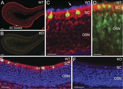
Fig. 7.
Strong immunoreactivity of muscarinic AChR subtype M3 in supporting cells of the MOE. A–D: immunolabeling results using the Research & Diagnostic (R&D) anti-M3 antibody. E and F: immunolabeling results using the Sigma anti-M3 antibody. A: low-magnification confocal image from a MOE section from a ChAT(BAC)-eGFP mouse showing strong M3 immunoreactivity (red) in the cell bodies of supporting cells (SC). WT, wild-type. B: confocal image from a MOE section of a mAChR M3 knockout (KO) mouse, showing absence of M3 immunoreaction. Sections in A and B were processed for immunoreaction and imaging under the same conditions. C: high-magnification image showing strong immunoreactivity for M3 (red) in the cell bodies of supporting cells in a MOE section of ChAT(BAC)-eGFP mouse. Some of their basal processes were also labeled. The cilia of OSNs were visualized using an antiacetylated tubulin (blue, pointed by an arrow). The M3 immunoreactivity is absent in the cilia. D: immunoreactivity of M3 (red) in a MOE section of a TRPM5-GFP mouse, showing the M3 antibody did not label the OSNs. The expression of GFP marks the TRPM5-expressing OSNs (Lin et al. 2007). E: confocal image obtained from a section labeled with the Sigma anti-M3 antibody, showing similar M3 immunoreaction in supporting cells as the R&D anti-M3 antibody. In most regions of the MOE, the Sigma anti-M3 antibody labeled only supporting cells. F: confocal image taken from a mAChR M3 knockout, showing abolished M3 immunoreactivity in supporting cells. Scale: A and B = 100 μm, C–F = 20 μm.

