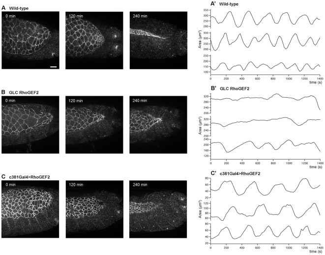
Figure 3. Loss and gain of function of DRhoGEF2 results in dorsal closure delay and impaired AS cell pulsations.
(A–C) Stills from movies during dorsal closure in embryos marked with Ubi-DECadherin-GFP. (A) Wild-type. (B) Maternal DRhoGEF2 mutants expressing Ubi-DECadherin-GFP. (C) Embryos marked with Ubi-DECadherin-GFP where UAS-DRhoGEF2 was overexpressed only in the AS cells. Embryos are shown at time 0, 120 and 240 min. Starting of dorsal closure (time 0) was considered when germ band was completely retracted. At 240 min, WT almost reach the end of dorsal closure, whereas DRhoGEF2 maternal mutants and c381GAL4/UAS-DRhoGEF2 holes are still open. Note that cell area is increased in DRhoGEF2 maternal mutants and decreased in c381GAL4/UAS-DRhoGEF2. The scale bar represents 20 µm. (A′–C′) Apical cell surface area oscillations of three representative AS cells from (A′) Wild-type, (B′) DRhoGEF2 maternal mutants, and (C′) c381GAL4/UAS-DRhoGEF2. Amplitude is in µm2 and time is in seconds (s). All AS cell pulsation analysis was performed on stage 13 embryos.

