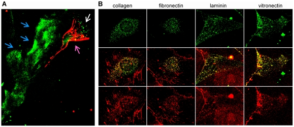
Figure 3. Co-internalization of SPARC and ECM proteins in fibroblasts.
(A) Primary mouseSPARC-null fibroblasts were attached to glass coverslips overnight and treated with red AF594-labeled collagen and green AF488-labeled SPARC. After 1 h of treatment, strong fibrillar collagennetworks were deposited at the trailing edge of a moving fibroblast (purple arrow). Fluorescent SPARC bound ECM tracks, deposited along the movement path (blue arrows) and was internalized at the leading edge of the fibroblast (white arrow). (B) SPARC-null fibroblasts were plated and treated with green AF488-labeled SPARC and red AF594-labeled ECM proteins as above. Allmatrix proteins were deposited into apparently normal extracellular networks and internalized by the cells. Strong co-localizationof all internalized ECM proteins with SPARC is evident by the yellow overlap of the two colors.

