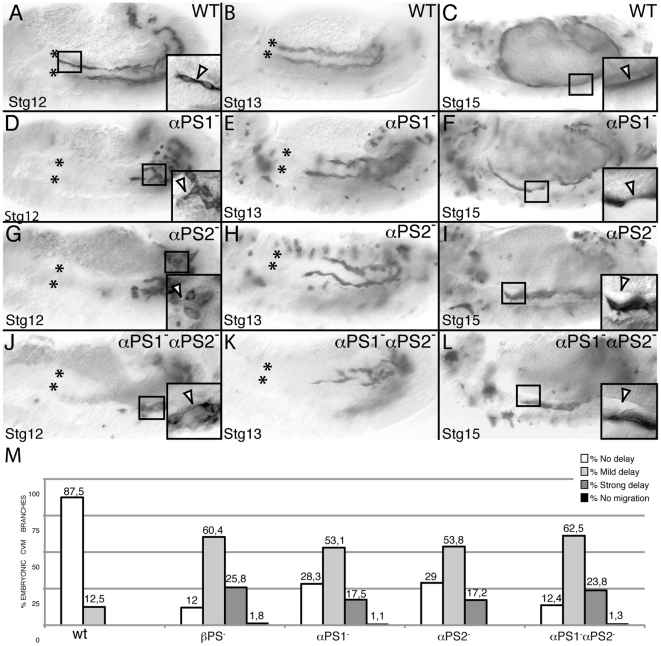
Figure 3. PS1 and PS2 integrins are required for proper CVM migration.
CVM cells are visualized using the combination G447.2-GAL4/UAS-CD2 and an anti-CD2 antibody staining. CVM cells from Stgs12 and 13 αPS1 (D, E, M) and αPS2 (G, H, M) mutant embryos are delayed in their migration as compared with wild type cells (A, B, M, yet they still send projections (D, G, arrowhead in magnification in black box) (F, I) In addition, CVM fibers from these mutant embryos detach from the vm at Stg15 (arrowhead in magnification in black box). (J–L) These phenotypes are enhanced in αPS1αPS2 double mutant embryos, phenocopying the defects observed in βPS mutant embryos. (M) Quantification of the CVM migration phenotype in Stg13 embryos of the designated genotypes.

