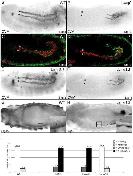
Figure 7. LamininW, but not lamininA, is required for CVM migration.
CVM cells are visualized using the combination 5053A-GAL4/UAS-srcGFP (A–D, F, G, H) or G447.2-GAL4/UAS-CD2 (E) and anti-GFP or anti-CD2 antibody staining, respectively. (A) Wild type embryo. (B) In absence of Laminin function, CVM cells fail to migrate. (C) During Stg12, wild type CVM cells (red) migrate in close contact with the vm, visualized with anti-FasIII antibody (green). However, Lamβ mutant CVM cells contact an intact vm but fail to migrate (D). (E) CVM migration is unaffected in Lamα3, 5 mutant embryos. (F) Conversely, CVM cells of Lamα1, 2 mutant embryos show a delay in their migration similar to that observed in Lamβ mutant embryos (B). (G, H) Attachment of CVM fibers to the vm is affected in stage 15 Lamα1, 2 mutant embryos (H) compare to wild type (G) (arrowhead in magnification in black box). (I) Quantification of the CVM migration phenotype in Stg13 embryos of the indicated genotypes.

