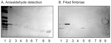
Figure 2. Binding of F4ad fimbriae to slow-migrating non-acid glycosphingolipid fractions isolated from chicken erythrocytes.
Chemical detection by anisaldehyde (A), and autoradiograms obtained by binding of 125I-labeled F4ad fimbriae (B). The lanes were: Lane 1, non-acid glycosphingolipids of chicken erythrocytes, 40 µg; Lanes 2–9, glycosphingolipid fractions isolated from chicken erythrocytes, 0.5–2 µg/lane.

