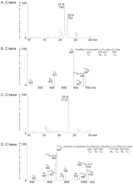
Figure 3. Characterization of the F4 fimbriae binding tetra- and hexaglycosylceramide of chicken erythrocytes.
(A) Base peak chromatogram from LC-ESI/MS of the saccharide obtained by digestion with Rhodococcus endoglycoceramidase II of the F4-binding glycosphingolipid fraction C:tetra-II from chicken erythrocytes. (B) MS2 spectrum of the ion at m/z 747 (retention time 21.4 min). (C) Base peak chromatogram from LC-ESI/MS of the saccharide obtained by digestion with Rhodococcus endoglycoceramidase II of the F4-binding glycosphingolipid fraction C:hexa from chicken erythrocytes. (D) MS2 spectrum of the ion at m/z 1112 (retention time 23.0 min).

