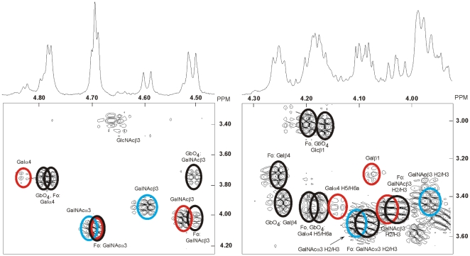
Figure 4. Proton NMR of the F4-binding glycosphingolipid (fraction C:tetra-I) from chicken erythrocytes.
Anomeric regions of the 600 MHz proton NMR of the F4-binding glycosphingolipid (fraction C:tetra-I) from chicken erythrocytes (30°C). The sample was dissolved in dimethyl sulfoxide-D2O (98∶2, by volume) after deuterium exchange. Below each section the corresponding DQF-COSY spectrum showing mainly the H1/H2 connectivities are displayed. Connectivities stemming from the same structure are color-coded by superimposed ellipses. Thus, the Forssman pentaglycosylceramide (Fo; GalNAcα3GalNAcß3Galα4Galß4Glcß1Cer) and globoside (GbO4; GalNAcß3Galα4Galß4Glcß1Cer) are black, whereas the two novel four-sugar compounds B and C are colored blue and red, respectively. Furthermore, only connectivities other than H1/H2 ones are denoted as such.

