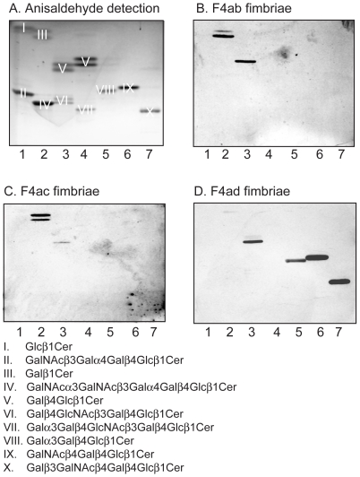
Figure 12. Binding of F4 fimbriae to reference glycosphingolipids.
Chemical detection by anisaldehyde (A), and autoradiograms obtained by binding of 125I-labeled F4ab fimbriae (B), F4ac fimbriae (C), and F4ad fimbriae (D). The lanes were: Lane 1, glucosylceramide (Glcß1Cer) of porcine kidney with d18:1/t18:0-16:0-24:0 ceramide, 4 µg, and globotetraosylceramide (GalNAcß3Galα4Galß4Glcß1Cer) of human erythrocytes with d18:1-16:0-24:0 ceramide, 4 µg; Lane 2, galactosylceramide (Galß1Cer) of bovine brain from Sigma-Aldrich with d18:1-h18:0-h24:0 ceramide, 4 µg, and Forssman pentaglycosylceramide (GalNAcα3GalNAcß3Galα4Galß4Glcß1Cer) of dog intestine with d18:1-16:0 and 24:0 ceramide, 4 µg; Lane 3, lactosylceramide (Galß4Glcß1Cer) with t18:0-h16:0-h24:0 ceramide of dog intestine, 4 µg, and neolactotetraosylceramide (Galß4GlcNAcß3Galß4Glcß1Cer) of human neutrophils d18:1-16:0 and 24:1 ceramide, 4 µg; Lane 4, lactosylceramide (Galß4Glcß1Cer) with d18:1-16:0-24:1 ceramide of human neutrophils, 4 µg, and B5 pentaglycosylceramide (Galα3Galß4GlcNAcß3Galß4Glcß1Cer) of rabbit erythrocytes with d18:1-16:0 and 24:0 ceramide, 4 µg; Lane 5, isoglobotriaosylceramide (Galα3Galß4Glcß1Cer) of cat intestine with t18:0-h22:0 and h24:0 ceramide, 4 µg; Lane 6, gangliotriaosylceramide (GalNAcß4Galß4Glcß1Cer) of guinea pig erythrocytes with d18:1-16:0 and 24:0 ceramide, 4 µg; Lane 7, gangliotetraosylceramide (Galß3GalNAcß4Galß4Glcß1Cer) of mouse intestine with t18:0-h16:0 and h24:0 ceramide, 4 µg. The glycosphingolipids visualized with anisaldehyde in (A) are marked with Roman numbers, and the corresponding glycosphingolipid structures are given below the chromatograms.

