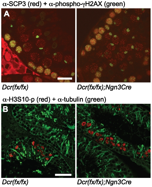
Figure 3. Meiotic analysis of Dicer1 knockout testes.
A) Confocal immunofluorescence microscopy of testis sections from control and knockout mice. An anti-SCP3 antibody (red) was used to detect synaptonemal complexes and an antibody against phosphorylated γH2AX (green) was used to visualize unsynapsed X and Y chromosomes in the sex body of pachytene spermatocytes. Scale bar: 20 µm. B) An antibody against phosphorylated H3 Serine 10 (red) was used to visualize meiotic metaphases. Double-staining with anti-tubulin (green) was used to visualize meiotic spindles and general organization of microtubular network in the seminiferous epithelium. Scale bar: 50 µm.

