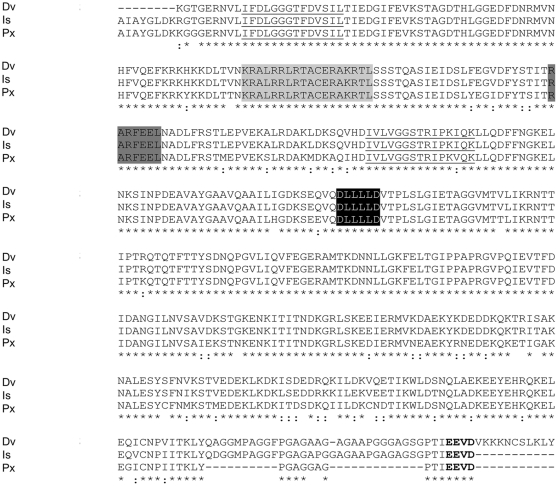
Figure 4. Multiple sequence alignments for heat shock protein from D. variabilis male transcriptome versus other species.
Multiple sequence alignments (ClustalW) of the deduced amino acid sequence of a putative D. variabilis Hsp70 with other putative Hsps from the Ixodida are compared. Dermacentor variabilis (Dv; contig11387), Ixodes scapularis (Is; EEC03688), Plutella xylostella (Px; BAE48743). Underlined residues show two of the three signature sequences of Hsp70 [78]. Residues shaded in light grey indicate one of the two bipartite nuclear localization signals common in Hsp70 [78]. Dark grey signifies the non-organellar consensus motif [79]. The linker region between ATPase and peptide domains is shaded in black [80]. The conserved EEVD motif at the C-terminus is bolded [78]. Asterisks denote identical residues, two dots signify conservative substitutions.

