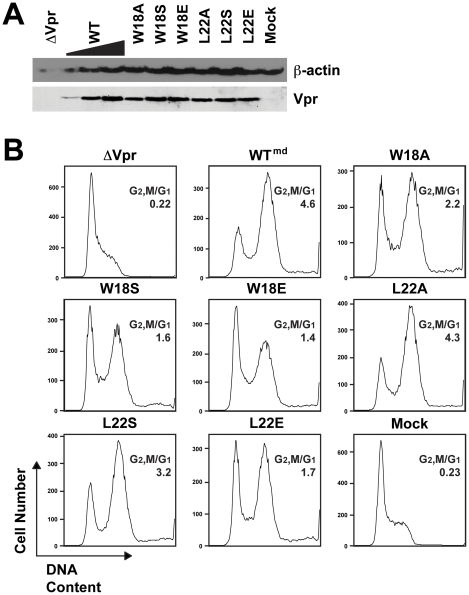
Figure 3. Hydrophobic residues on Vpr helix-1 are important for virion-delivered Vpr cell cycle arrest.
WT or mutant Vpr proteins (denoted by the single letter amino acid changes) were delivered (Vprv) into Jurkat cells. Virions containing WT Vpr were titrated (md, medium) so that a matched Vpr protein control could be compared to the mutants. (A) Western blot of the Jurkat cells for WT and mutant virion-delivered Vpr (bottom). β-actin is shown as a protein loading control (top). (B) Histograms of cell cycle analysis at 41 hr post-infection show DNA content of PI-stained cells by flow cytometry. All samples represent 10,000 cellular events. G1 and G2/M populations were modeled using the Watson Pragmatic cell cycle model, and the G2,M/G1 ratio in each infection is shown. The data are representative of three experiments.

