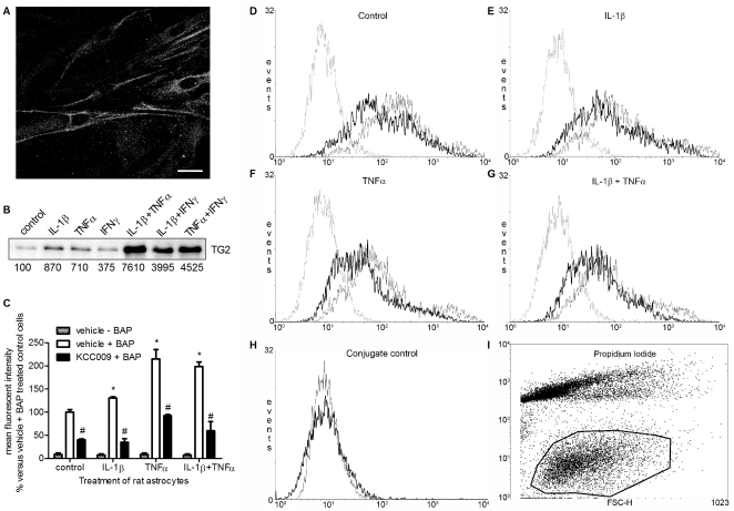
Figure 2. TG2 is expressed on the surface of rat astrocytes.
A) Life non-permeabilized cells were immunofluorescently labeled to detect TG2 on the cell surface of rat astrocytes. Scale bar: 20 µm B) Cell surface-biotinylated proteins were immunoprecipitated, separated by SDS-PAGE, and immunoblotted for TG2. Bands were semi-quantified and expressed as % compared to control. C) FACS analysis of BAP incorporation on the surface of control rat astrocytes or astrocytes treated with IL-1β, TNFα or IL-1β+TNFα in the absence or presence of KCC009. Data represent mean fluorescent intensity from 3 independent experiments. *P<0.05 versus vehicle-treated control cells, #P<0.05 versus vehicle-treated control or matched cytokine-treated. D–G) FACS plots of BAP incorporation on the surface of rat astrocytes D) untreated or treated with E) IL-1β, F) TNFα or G) IL-1β+TNFα and subsequent inhibition of TG2 activity. Dotted line: vehicle − BAP treated astrocytes; grey line: vehicle + BAP treated astrocytes; black line: KCC009 + BAP treated astrocytes H) Effect of conjugate (avidin-Alexa Fluor-488). Grey line: untreated cells –BAP−avidine-Alexa Fluor, black line: untreated cells –BAP+avidine-Alexa Fluor 488. I) Encircled astrocytes which were PI negative were gated for BAP.

