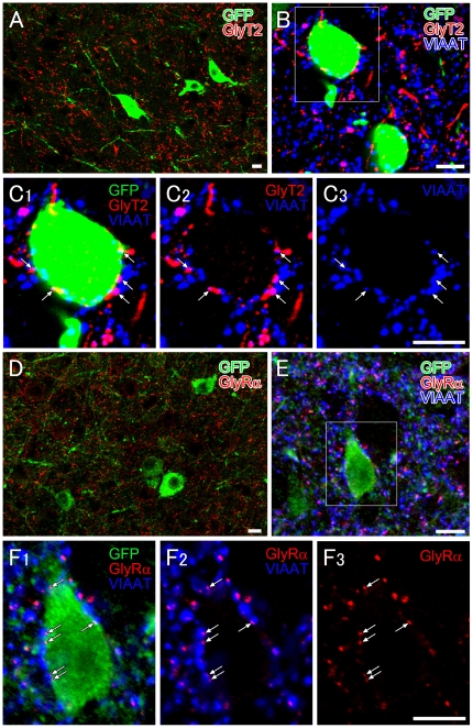
Figure 5. Immunofluorescence showing glycinergic innervations to orexin neurons.
A. Double immunofluorescence showing distribution of GlyT2-positive glycinergic varicose fibers (red) and GFP-positive orexin neurons (green) in the LHA. B, C. Triple immunofluorescence showing that orexin neurons (GFP, green) are associated with GlyT2-positive (red)/VIAAT-positive (blue) varicosities (arrows). D. Double immunofluorescence showing the distribution of GlyRα- (red) and GFP-positive orexin neurons (green) in the LHA. E, F. Orexin neurons (green) display numerous VIAAT-positive inhibitory terminals (blue), some of which are associated with GlyRα immunoreactivity (red) on the surface of orexin neurons. Scale bars, 10 µm.

