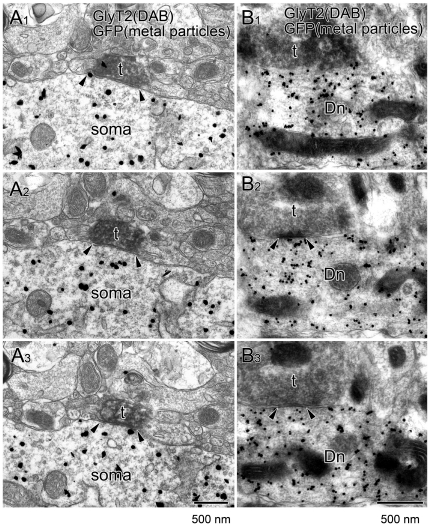
Figure 6. Immunoelectron microscopy showing glycinergic synapse formation on orexin neurons.
Consecutive images from double-labeling preembedding immunoelectron microscopy for GFP (silver-intensified immunogold) and GlyT2 (immunoperoxidase). Note that a GlyT2-positive terminal (filled with diffuse DAB precipitates) makes a symmetrical synaptic junction (arrowheads) onto the soma (A) and dendritic shaft (B) of a GFP-positive orexin neuron. Scale bar, 500 nm.

