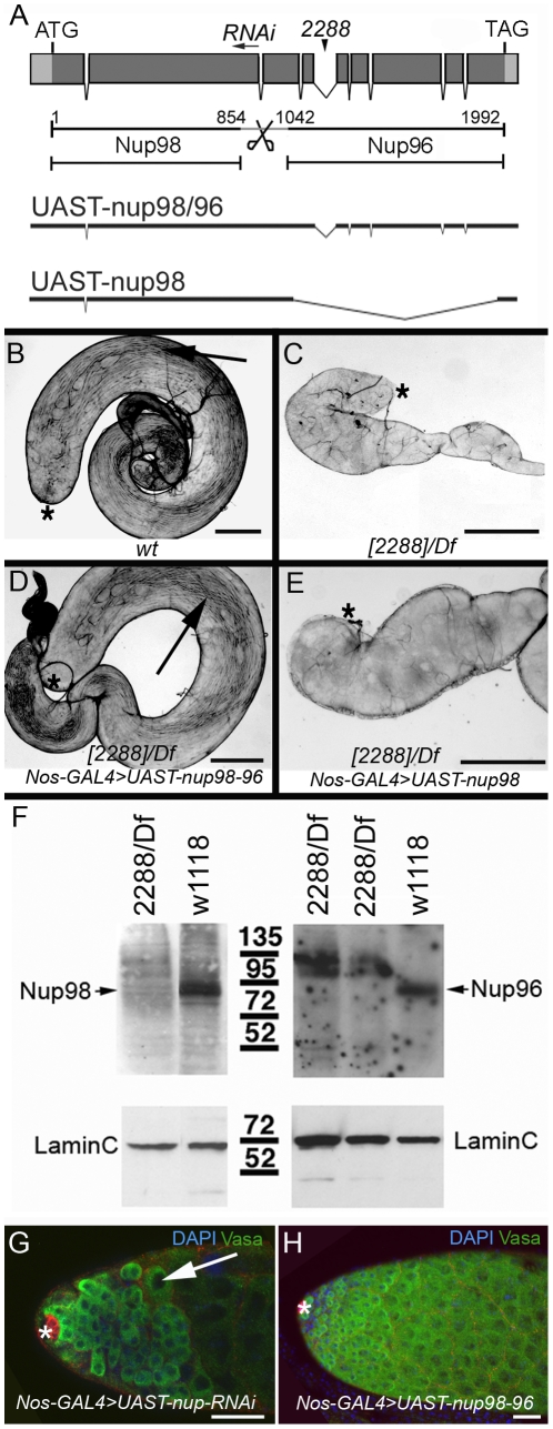
Figure 3. Mutations in nup98-96 disrupt gametogenesis.
(A) Top: Intron-exon structure of the nup98-96 locus. Coding region in dark grey. Gene products as indicated. Bottom: Rescue constructs containing the whole transcription unit for nup98-96 or nup98 only. Arrowhead: Pogo-insertion in nup98-962288, arrow: RNAi-sequences. (B-E) Bright field images of whole testes. Genotypes as indicated. Arrows point to sperm. Asterisks mark the apical tips. Scale bars: 100 µm. (F) Western blot analysis. Genotypes and antibodies as indicated. The Nup96 antibody does not detect a 95 kd protein in extracts from mutant animals. Instead, the Nup96 antibody detects a high molecular weight bands that may be abnormal Nup96 protein or Nup98-Nup96 polyprotein. The Nup98 antibody detects extremely low levels a 95 kd protein in the mutant compared to the control. (G, H) Apical tips of adult testes with germ line expression of (G) UAST-nup98-96-RNAi (arrow points to a single spermatocyte), and (H) UAST-nup98-96. Asterisks mark the apical tips. Scale bars: 30 µm.

