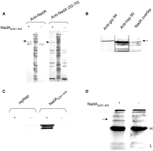
Figure 1. NadAΔ351–405 binds to human hsp90 in an overlay-assay.
A) Samples of total monocytes extracts obtained with 1% TX-100 were separated by SDS-PAGE and challenged or not as indicated with 1 µM NadAΔ351–405 after blotting on nitrocellulose. After extensive washings, the polypeptide bands able to retain NadA were detected with antibodies to the whole adhesin or to NadA 52–70, as indicated. The picture is from a representative experiment out of four and arrows point to the NadA-interacting ∼90 KDa protein. B) Monocytes detergent extracts prepared as in A) were separated by SDS-PAGE, blotted on nitrocellulose and subjected to western blot with anti- grp94 and hsp90 antibodies or to an overlay assay with NadA/anti-NadA antibodies. The mobility of the NadA interacting protein and of hsp90 or grp94 was compared. C) Purified human hsp90 were subjected to overlay assay with (1 µM) or without NadAΔ351–405 and HP-NAP as indicated. D) Whole TX-100 extracts from human monocytes were incubated with protein A sepharose-bound anti-NadA IgG in the presence or not of NadAΔ351–405. After washings, beads were treated with LSB containing 4%SDS and 10% βme and analyzed by western blot after SDS-PAGE using anti-hsp90 antibodies and alkaline phosphatase-conjugated anti-IgG secondary antibodies. Arrow points to hsp90, H and L indicate antibody heavy and light chains respectively.

