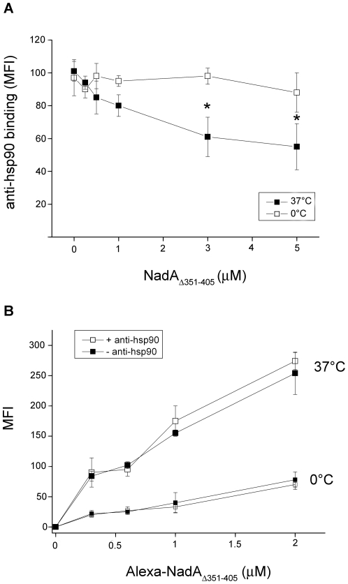
Figure 4. Analysis of the reciprocal interference of NadAΔ351–405 and anti-hsp90 antibodies to the monocyte surface at 37° and 0°C.
A) Human monocytes were incubated for 1 hours at 0°C in RPMI, 10% FCS, containing NadAΔ351–405 at the indicated concentrations. After washing at 0°C cells were further incubated with anti-hsp90 antibody and anti-IgG PE-labeled secondary antibodies. MFI was then analyzed by flow-cytofluorimetry. Data are from an experiment representative of four, and bars represent +/− SE. Asterisk indicates signals significantly different (p<0.05) from control (0°C). B) Monocytes were pre-treated or not with anti-hsp90 antibodies (40 µg/ml) for 1 hour at 0°C, washed as above and further incubated at the indicated temperature with Alexa-labeled NadAΔ351–405. Graphs are made with mean values from a representative experiment of four. Bars are +/− SE.

