
Figure 5.
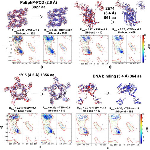
Application of TOP to different protein structures. For PaBphP-PCD, the RamaMap for four of the eight chains is shown. For the DNA-binding protein and cytochrome b6f membrane complex (2E74) structures, the DNA and a few cofactors are part of the crystal structure but are not displayed.