
Figure 2.
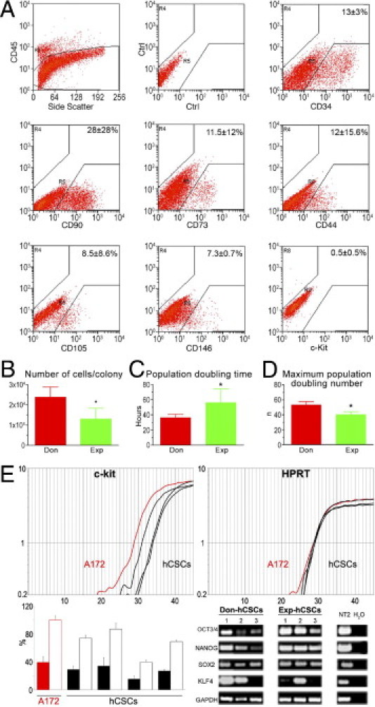
Properties of hCSCs. A: Surface phenotype of small cardiac cells isolated from donor and explanted hearts. B–D: Growth properties of hCSCs. Data are reported as means ± SD. *P < 0.05 versus Don. E: Representative amplification curves of c-Kit and HPRT (housekeeping gene) expression in cell line A172 (red), which was used as positive control and in hCSC samples (black). In the histogram, solid bars indicate the relative expression of c-Kit in A172 cells (red) and in hCSCs (black), and open bars indicate the expression of c-Kit in hCSCs normalized versus A172 cells. Transcripts for OCT3/4, NANOG, SOX2, and KLF4 are present in hCSCs obtained from donor (Don-hCSCs) and explanted (Exp-hCSCs) hearts. The neuronally committed human teratocarcinoma cell line NT2 was used as positive control. PCR products were run on 1.8% agarose gel.