
Figure 7.
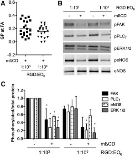
Membrane order regulates VEGF signaling. (A) Endothelial cells that adhered to 1:103 RGD/EO6 and 1:106 RGD/EO6 surfaces for 3 h were cholesterol-depleted with 10 mM mβCD for 25 min. Cholesterol depletion significantly decreased membrane order at FAs in cells plated on either surface, such that the mean GP values of FAs of cholesterol-depleted cells (indicated by the horizontal bar) are not significantly different. (B) Immunoblots for the phosphorylation of FAK, PLCγ, ERK1/2, and eNOS for control and cholesterol-depleted cells treated with 25 ng/ml VEGF for 10 min. Probing for nonphosphorylated proteins, as shown for eNOS, was used as loading control. (C) Quantification of VEGF-induced signaling activity expressed as the ratio of phosphorylated/total protein, and normalizing the ratio to cell lysates from 1:103 RGD/EO6 surfaces. Data and error bars represent the mean ± SD of three independent experiments; ∗p < 0.05 relative to non-mβCD-treated cells on equivalent surfaces. Signaling activity in cholesterol-depleted cells on 1:103 RGD/EO6 versus 1:106 RGD/EO6 is not significantly different.