
Figure 1.
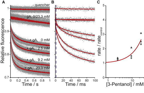
3-Pentanol's bilayer-perturbing effect, as determined using a gA-based fluorescence assay. Normalized fluorescence time courses for fluorophore-loaded LUVs incubated with varying [3-pentanol], with and without 260 nM gA and with and without external quencher. (A) Average traces (lines) and results from all repeats (>7 per condition; dots) over 1 s. (B) One repeat for each experimental condition in A (dots), as well as stretched exponential fits (2–100 ms) to those repeats (solid lines). The stippled line denotes the 2 ms mark. (C) The normalized quenching rate relative to rates of vesicles with the same amount of gA and no added 3-pentanol. Results are for three different days of experiments (differently shaped symbols); the solid line indicates a f([alc]) = 1 + [alc]/D fit to the results.