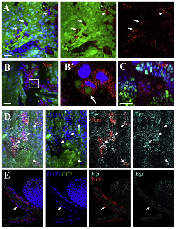
Figure 3. Associated Hemocytes Express Eiger.
In all panels, GFP-labeled clones (green) of RasV12; scrib−/− cells were created in developing eye-antennae discs in egr+/+ hosts, except in (C) where the host was egr−/−. DAPI labeled nuclei are shown in blue. (A) Anti-Eiger (red) in RasV12; scrib−/− clones indicated that some tumor-associated cells express Eiger (arrows). (B and C) Anti-Nimrod C1 staining (Nim, red) labeled tumor-associated hemocytes. (B′) shows a high magnification view from the boxed area in (B). The arrow points to a phagocytosed tumor cell fragment. (D and E) Anti-Nimrod C1 (red) and Anti-Eiger (cyan) staining from a tumor (D) and a trachea branch (E) from the same animal. Left panels are merged images; the other panels show stains as labeled. Arrows point to hemocytes. Scale bars = (A), (B), (D), and (E), 20 μm, and (C), 25 μm. See also Figure S7.

