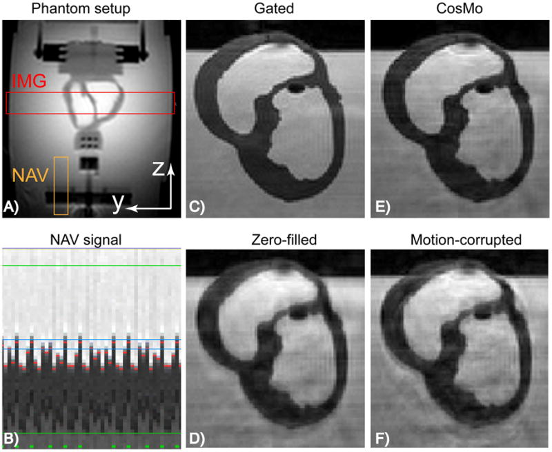
Figure 2.

Non-rigid motion phantom: A) 2D image of the phantom setup demonstrating position of the imaging slab and NAV. B) NAV signal tracking the heart and acquiring the inner k-space lines within 5 mm gating window (after this stage the gating window is increased to acquire the rest of k-space lines). Green, red, and blue lines represent the accepted k-space segments, measured phantom displacements, and gating window size, respectively. C) Prospectively gated NAV with 5mm gating window. D) Zero-filled image, where the outer k-space data acquired outside the gating window are replaced with zero. E) CosMo reconstructed image using 51% k-space data. F) Motion-corrupted image, where the outer k-space data acquired outside the gating window are used in the image reconstruction.
