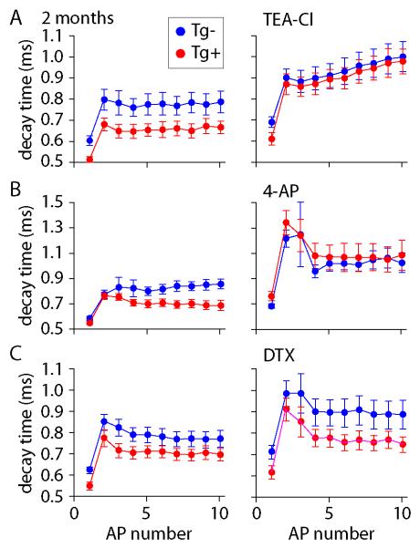
Figure 6. Pharmacological blockade of difference in AP waveform.
(A-C) Effects of potassium channel blockers on the 90-10% decay times of action potentials in 2 month-old CRND8 mice. Decay times are reported for trains for 10 APs. Each symbol is the mean ± SEM for Tg− (blue) or Tg+ mice (red). In each recording decay times were measured first under control conditions (left) and the after application of blocker (right). (A) Effect of 300 μM tetraethylammonium chloride (TEA-Cl); n = 5 Tg− and 6 Tg+ neurons. Decay times were significantly different under control conditions (P<0.05) and this difference was eliminated by TEA (P>0.05). (B) Effect of 100 μM 4-aminopyridine (4-AP); n = 7 Tg− and 6 Tg+ neurons. Decay times were significantly different under control conditions (P<0.05) and this difference was eliminated by 4-AP (P>0.05). (C) No effect of 100 nM dendrotoxin (DTX); n = 10 Tg− and 10 Tg+ neurons. Decay times were significantly different under control conditions (P<0.05) and in DTX (P<0.05).

