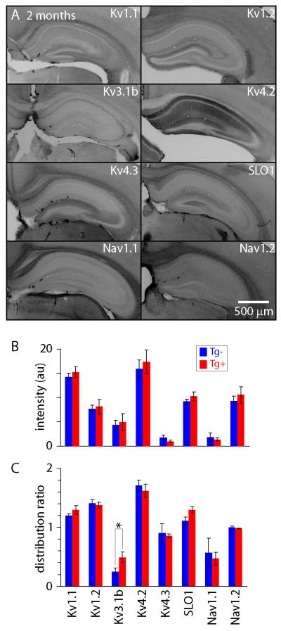
Figure 7. Channel immunohistochemistry in 2 month-old CRND8 mice.
(A) Widefield images of hippocampi from 2 month-old Tg+ mice stained for several potassium and sodium channels. Intensities were inverted for display purposes. (B) Fluorescence intensities in CA1 (arbitrary intensity units). Intensities were averaged across stratum oriens, stratum pyramidale and stratum radiatum. There was no significant difference in intensity between 2 month-old Tg+ and Tg− mice immunostained with any of the anti-bodies tested. Each bar is the mean (+/− SEM) of 3-7 mice, with 3-6 sections from each mouse. (C) Immuofluorescence intensities in straum oriens and radiatum normalized to the intensity in stratum pyramidale in CA1. Fluorescence intensity differed between 2 month-old Tg+ and Tg− mice only for Kv3.1b (asterisk denotes P<0.05, unpaired t-test).

