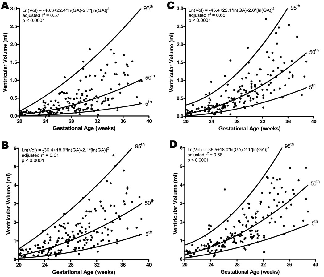
Figure 1.
Volume measurements taken in end systole and end diastole for the left and right ventricles increased significantly with advancing gestational age (A: left ventricle in end systole, B: left ventricle in end diastole, C: right ventricle in end systole, D: right ventricle in end diastole). The regression line with the 5% and 95% confidence intervals is plotted with the regression equation, adjusted r2, and p value for each. Additionally, the median ventricular volumes were significantly greater for the right side in both systole (Right: 0.50 ml, IQR: 0.2 – 0.9; Left: 0.27 ml, IQR: 0.1 – 0.5; p<0.001) and diastole (Right: 1.20 ml, IQR: 0.7 – 2.2; Left: 1.03 ml, IQR: 0.5 – 1.7; p<0.001).

