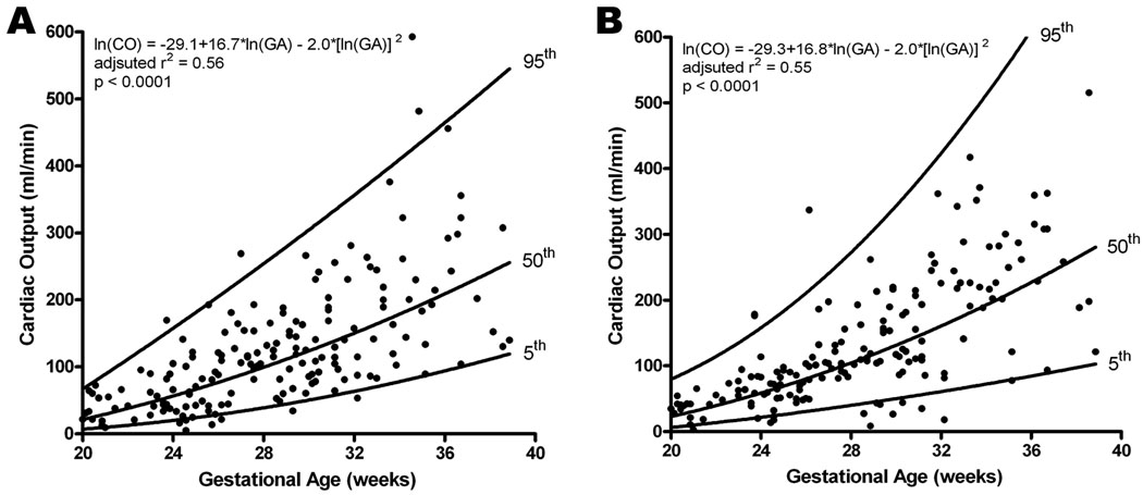
Figure 3.
Cardiac output (CO; stroke volume x fetal heart rate) obtained for left (A) and right (B) ventricles increased with advancing gestational age; however, there was no significant difference found between the two ventricles. The regression line with the 5% and 95% CI’s is plotted with the regression equation, adjusted r2, and p value for each.

