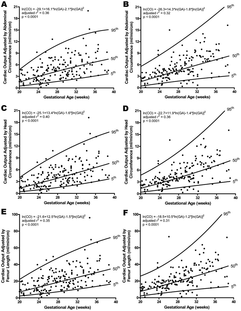
Figure 5.
Cardiac output (CO; stroke volume x fetal heart rate) obtained for left (A, C, E) and right (B, D, F) ventricles, adjusted by fetal biometric measurement, increased significantly with advancing gestational age. There was no significant difference when left and right were compared. The regression line with the 5% and 95% CI’s is plotted with the regression equation, adjusted r2, and p value for each.

