
Figure 4.
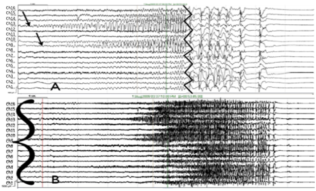
(A) Focal onset right occipital seizure (arrows) with secondary generalization on iEEG, onset and termination. Channel labels are 16 at top down to 1 at bottom). Jagged vertical line denotes invervening portion of record (removed) showing secondarily generalization. (B) Regional onset seizure (sharp wave to left of red line defines onset time, bracket denotes channels involved). Event begins with diffuse electrodecrement and widespread low voltage fast activity prominent in channels 15, 16, 7, 8, 4 and 1, among others. At these filter settings and in this montage, the seizure cannot be better localized.