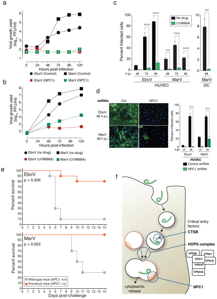
Figure 4. NPC1 function is required for infection by authentic Ebola and Marburg viruses.
a, NPC1 patient fibroblasts were exposed to EboV or MarV at a multiplicity of infection (MOI) of 0.1. Supernatants were harvested and yields of infectious virus were measured. *below detection limit. b, Vero cells treated with DMSO or U18666A (20 μM) were infected with EboV or MarV at an MOI of 0.1 and yields of infectious virus were measured. c, Human peripheral blood monocyte-derived dendritic cells (DC) and umbilical-vein endothelial cells (HUVEC) were infected in the presence or absence of U18666A at an MOI of 3 and the percentage of infected cells was determined by immunostaining. d, HUVEC were transduced with lentiviral vectors expressing a non-targeting shRNA (Ctrl) or an shRNA targeting NPC1, infected with EboV or MarV at an MOI of 3 and the percentage of infected cells was determined. Representative images of cells are also shown: green, viral antigen; blue, nuclear counterstain. For panels a–d, Means ±SD are shown (n=2 to 3). In panels a–b, error bars are not visible because they are within the symbols. For panels c–d, ** p-value < 0.01, *** p-value < 0.001. e, Survival of NPC1+/+ and NPC1−/+ mice (n=10 for each group) inoculated i.p. with ~1000 pfu of mouse-adapted EboV or MarV. f, A proposed hypothetical model for the roles of CatB, the HOPS complex, and NPC1 in Ebola virus entry.

