
Figure 4.
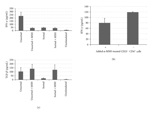
The effects of α-MSH on CD25− CD4+ T cells. The CD4 T cells were isolated from the draining lymph node as in Figure 1 and were stained with CD25 antibody. The stained cells were sorted, and the CD25− cells were placed in culture, stimulated, and treated with α-MSH. (a) After 48 hours, the supernatant was assayed for IFN-γ and TGF-β. The sorted CD25− CD4+ T cells (Sorted) did not produce IFN-γ or TGF-β; however, when treated with α-MSH (Sorted + MSH) did produce TGF-β but not IFN-γ. (b) The α-MSH-treated CD25− CD4+ T cells were transferred to cultures of activated Th1 cells. After 48 hours of incubation IFN-γ was measured in the supernatant. There was no statistical difference in IFN-γ production by the Th1 cells in culture with or without α-MSH-treated CD25− CD4+ T cells.