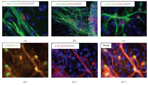
Figure 3.
Immunofluorescence analysis of Lbx1 expression in ES cell-derived cells. (a) Sarcomeric α-actinin- (green)-labeled skeletal muscle cell progenitors partially costained for Lbx1 (red) at day 5 + 14 of spontaneous ES cell differentiation. (b) β III tubulin- (green)-positive neuronal cells partially coexpressed Lbx1 (red) at day 5 + 11. (c) Lbx1 staining (red) was clearly located to the nuclear region of β III tubulin- (green)-positive neurons. Triple immunofluorescence staining revealed the coexpression of (d′) Lbx1 (green) and GABA (orange) in (d′′) β III tubulin- (dark red)-positive neurons. (d′′′) Merged image. Nuclei were labeled with fluorescent marker DAPI (blue). Bar = 20 μm.

